Exploring transformer reliability in clinically significant prostate cancer segmentation: A comprehensive in-depth investigation
IF 5.4
2区 医学
Q1 ENGINEERING, BIOMEDICAL
Computerized Medical Imaging and Graphics
Pub Date : 2024-11-17
DOI:10.1016/j.compmedimag.2024.102459
引用次数: 0
Abstract
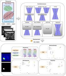
Despite the growing prominence of transformers in medical image segmentation, their application to clinically significant prostate cancer (csPCa) has been overlooked. Minimal attention has been paid to domain shift analysis and uncertainty assessment, critical for safely implementing computer-aided diagnosis (CAD) systems. Domain shift in medical imagery refers to differences between the data used to train a model and the data evaluated later, arising from variations in imaging equipment, protocols, patient populations, and acquisition noise. While recent models enhance in-domain performance, areas such as robustness and uncertainty estimation in out-of-domain distributions have received limited investigation, creating indecisiveness about model reliability. In contrast, our study addresses csPCa at voxel, lesion, and image levels, investigating models from traditional U-Net to cutting-edge transformers. We focus on four key points: robustness, calibration, out-of-distribution (OOD), and misclassification detection (MD). Findings show that transformer-based models exhibit enhanced robustness at image and lesion levels, both in and out of domain. However, this improvement is not fully translated to the voxel level, where Convolutional Neural Networks (CNNs) outperform in most robustness metrics. Regarding uncertainty, hybrid transformers and transformer encoders performed better, but this trend depends on misclassification or out-of-distribution tasks.

探索具有临床意义的前列腺癌分段中变压器的可靠性:全面深入的调查
尽管变换器在医学图像分割中的作用日益突出,但其在具有临床意义的前列腺癌(csPCa)中的应用却一直被忽视。人们对域偏移分析和不确定性评估的关注极少,而这对安全实施计算机辅助诊断(CAD)系统至关重要。医学影像中的域偏移指的是用于训练模型的数据与随后评估的数据之间的差异,这种差异是由成像设备、协议、患者群体和采集噪声的变化引起的。虽然最近的模型提高了域内性能,但对域外分布的鲁棒性和不确定性估计等领域的研究却很有限,导致对模型的可靠性举棋不定。相比之下,我们的研究涉及体素、病灶和图像层面的 csPCa,研究了从传统 U-Net 到尖端变换器的各种模型。我们重点关注四个关键点:稳健性、校准、分布外 (OOD) 和误分类检测 (MD)。研究结果表明,基于变换器的模型在图像和病变水平上表现出更强的鲁棒性,无论是在域内还是域外。然而,这种改进并没有完全转化到体素层面,在体素层面,卷积神经网络(CNN)在大多数鲁棒性指标上都表现出色。在不确定性方面,混合变换器和变换器编码器表现更好,但这一趋势取决于误分类或分布外任务。
本文章由计算机程序翻译,如有差异,请以英文原文为准。
求助全文
约1分钟内获得全文
求助全文
来源期刊
CiteScore
10.70
自引率
3.50%
发文量
71
审稿时长
26 days
期刊介绍:
The purpose of the journal Computerized Medical Imaging and Graphics is to act as a source for the exchange of research results concerning algorithmic advances, development, and application of digital imaging in disease detection, diagnosis, intervention, prevention, precision medicine, and population health. Included in the journal will be articles on novel computerized imaging or visualization techniques, including artificial intelligence and machine learning, augmented reality for surgical planning and guidance, big biomedical data visualization, computer-aided diagnosis, computerized-robotic surgery, image-guided therapy, imaging scanning and reconstruction, mobile and tele-imaging, radiomics, and imaging integration and modeling with other information relevant to digital health. The types of biomedical imaging include: magnetic resonance, computed tomography, ultrasound, nuclear medicine, X-ray, microwave, optical and multi-photon microscopy, video and sensory imaging, and the convergence of biomedical images with other non-imaging datasets.

 求助内容:
求助内容: 应助结果提醒方式:


