Deep learning-driven feature engineering for lung disease classification through electrical impedance tomography imaging
IF 4.9
2区 医学
Q1 ENGINEERING, BIOMEDICAL
引用次数: 0
Abstract
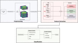
Thoracic imaging is vital for diagnosing lung diseases, as it provides a detailed visualization of the lungs. Despite significant advancements in medical imaging techniques, These methods pose critical challenges, such as high costs and the use of radiation in certain devices, which can raise serious concerns, limit accessibility, and increase potential health risks. Therefore main aim of this study addressing these issues by utilizing electrical impedance tomography (EIT), which is a non-invasive imaging technique that mitigates the risks of radiation exposure, reduces costs, and simplifies the interpretation of complex lung disease related patterns seen in traditional imaging methods. As EIT emerges as a promising imaging technique, this study investigates and develops a deep learning-based framework for classifying lung diseases using reconstructed EIT images. The proposed framework includes three feature extraction methods: Initial-Pretrained Weights Models (ResNet-50 and DenseNet-201), fine-tuned convolutional 3D networks, and fine-tuned convolutional 3D accompanied by dense layer networks. Various machine models fed by extracted features were employed for lung sound disease classification both as individual learners and ensemble classifiers. The framework was evaluated on three classification tasks: binary classification (healthy vs. non-healthy) achieving 89.55% accuracy, 3-class classification (obstructive-related, restrictive-related, and healthy) achieving 55.29% accuracy, and 5-class classification (asthma, chronic obstructive pulmonary disease, interstitial lung disease, pulmonary infection, and healthy) achieving 44.54% accuracy. The proposed methods outperform state-of-the-art results and introduce novel approaches to EIT imaging classification.

通过电阻抗断层成像进行肺病分类的深度学习驱动特征工程
胸部成像可提供肺部的详细图像,对诊断肺部疾病至关重要。尽管医学成像技术取得了长足的进步,但这些方法也带来了严峻的挑战,如高昂的成本和某些设备中辐射的使用,这些都会引起严重的担忧,限制了可及性,并增加了潜在的健康风险。因此,本研究的主要目的是利用电阻抗断层扫描(EIT)来解决这些问题,EIT 是一种非侵入性成像技术,可降低辐射风险、降低成本,并简化对传统成像方法中出现的复杂肺部疾病相关模式的解释。随着 EIT 成为一种前景广阔的成像技术,本研究调查并开发了一种基于深度学习的框架,用于利用重建的 EIT 图像对肺部疾病进行分类。所提出的框架包括三种特征提取方法:初始预训练加权模型(ResNet-50 和 DenseNet-201)、微调卷积三维网络和微调卷积三维伴密集层网络。在肺部疾病分类中,采用了以提取的特征为反馈的各种机器模型,既可以作为单个学习器,也可以作为集合分类器。该框架在三个分类任务中进行了评估:二元分类(健康与非健康)准确率达到 89.55%,三类分类(阻塞性相关、限制性相关和健康)准确率达到 55.29%,五类分类(哮喘、慢性阻塞性肺病、间质性肺病、肺部感染和健康)准确率达到 44.54%。所提出的方法优于最先进的结果,并为 EIT 成像分类引入了新方法。
本文章由计算机程序翻译,如有差异,请以英文原文为准。
求助全文
约1分钟内获得全文
求助全文
来源期刊

Biomedical Signal Processing and Control
工程技术-工程:生物医学
CiteScore
9.80
自引率
13.70%
发文量
822
审稿时长
4 months
期刊介绍:
Biomedical Signal Processing and Control aims to provide a cross-disciplinary international forum for the interchange of information on research in the measurement and analysis of signals and images in clinical medicine and the biological sciences. Emphasis is placed on contributions dealing with the practical, applications-led research on the use of methods and devices in clinical diagnosis, patient monitoring and management.
Biomedical Signal Processing and Control reflects the main areas in which these methods are being used and developed at the interface of both engineering and clinical science. The scope of the journal is defined to include relevant review papers, technical notes, short communications and letters. Tutorial papers and special issues will also be published.
 求助内容:
求助内容: 应助结果提醒方式:


