Estrogen replacement therapy reverses spatial memory loss and pyramidal cell neurodegeneration in the prefrontal cortex of lead-exposed ovariectomized Wistar rats
IF 2.9
Q2 TOXICOLOGY
引用次数: 0
Abstract
Background
Although menopause is a component of chronological aging, it may be induced by exposure to heavy metals like lead. Interestingly, lead exposure, just like the postmenopausal state, has been associated with spatial memory loss and neurodegeneration; however, the impact of hormone replacement therapy (HRT) on menopause and lead-induced spatial memory loss and neurodegeneration is yet to be reported.
Aim
The present study investigated the effect and associated mechanism of HRT on ovariectomized-driven menopausal state and lead exposure-induced spatial memory loss and neurodegeneration.
Materials and methods
Thirty adult female Wistar rats were randomized into 6 groups (n = 5 rats/group); the sham-operated vehicle-treated, ovariectomized (OVX), OVX + HRT, lead-exposed, OVX + lead, and OVX + Lead + HRT groups. Treatment was daily via gavage and lasted for 28 days.
Results
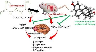
Ovariectomy and lead exposure impaired spatial memory deficit evidenced by a significant reduction in novel arm entry, time spent in the novel arm, alternation, time exploring novel and familiar objects, and discrimination index. These findings were accompanied by a marked distortion in the histology of the prefrontal cortex, and a decline in serum dopamine level and pyramidal neurons. In addition, ovariectomy and lead exposure induced metabolic disruption (as depicted by a marked rise in lactate level and lactate dehydrogenase and creatinine kinase activities), oxidative stress (evidenced by a significant increase in MDA level, and decrease in GSH level, and SOD and catalase activities), inflammation (as shown by significant upregulation of myeloperoxidase activity, and TNF-α and IL-1β), and apoptosis (evidenced by a rise in caspase 3 activity) of the prefrontal cortex. The observed biochemical and histological perturbations were attenuated by HRT.
Conclusions
This study revealed that HRT attenuated ovariectomy and lead-exposure-induced spatial memory deficit and pyramidal neurodegeneration by suppressing oxidative stress, inflammation, and apoptosis of the prefrontal cortex.

雌激素替代疗法可逆转铅暴露卵巢切除 Wistar 大鼠前额叶皮层的空间记忆丧失和锥体细胞神经退行性变
背景虽然更年期是自然衰老的一个组成部分,但它也可能是由铅等重金属暴露诱发的。有趣的是,铅暴露与绝经后状态一样,与空间记忆丧失和神经变性有关;然而,激素替代疗法(HRT)对绝经和铅诱导的空间记忆丧失和神经变性的影响尚未见报道。材料和方法将 30 只成年雌性 Wistar 大鼠随机分为 6 组(n = 5 只/组):假手术载体处理组、卵巢切除(OVX)组、OVX + HRT 组、铅暴露组、OVX + 铅组和 OVX + 铅 + HRT 组。结果卵巢切除术和铅暴露损害了空间记忆缺陷,表现为新手臂进入、新手臂停留时间、交替、探索新物体和熟悉物体的时间以及辨别指数显著减少。伴随这些发现的是前额叶皮层组织学的明显扭曲、血清多巴胺水平和锥体神经元的下降。此外,卵巢切除术和铅暴露还诱发了代谢紊乱(表现为乳酸水平、乳酸脱氢酶和肌酸激酶活性明显升高)、氧化应激(表现为 MDA 水平显著升高、GSH 水平显著降低、血清多巴胺水平显著升高和锥体神经元水平显著降低)、前额叶皮质的氧化应激(表现为 MDA 含量明显增加、GSH 含量减少、SOD 和过氧化氢酶活性降低)、炎症(表现为髓过氧化物酶活性、TNF-α 和 IL-1β 明显上调)和细胞凋亡(表现为 Caspase 3 活性升高)。结论这项研究表明,通过抑制氧化应激、炎症和前额叶皮质细胞凋亡,HRT 可减轻卵巢切除术和铅暴露引起的空间记忆缺失和锥体神经变性。
本文章由计算机程序翻译,如有差异,请以英文原文为准。
求助全文
约1分钟内获得全文
求助全文
来源期刊

Current Research in Toxicology
Environmental Science-Health, Toxicology and Mutagenesis
CiteScore
4.70
自引率
3.00%
发文量
33
审稿时长
82 days
 求助内容:
求助内容: 应助结果提醒方式:


