Flaring up the mesentery: Applying the LODOX eXero-DR® statscan for expedited postmortem angiography.

IF 0.8 Q4 RADIOLOGY, NUCLEAR MEDICINE & MEDICAL IMAGING
Zandré Smith , Elrinda de Bruin , Ilze Mentoor , Janette Verster
{"title":"Flaring up the mesentery: Applying the LODOX eXero-DR® statscan for expedited postmortem angiography.","authors":"Zandré Smith ,&nbsp;Elrinda de Bruin ,&nbsp;Ilze Mentoor ,&nbsp;Janette Verster","doi":"10.1016/j.fri.2024.200614","DOIUrl":null,"url":null,"abstract":"<div><h3>Rationale</h3><div>The affordable LODOX eXero-DR® statscan aids Forensic Pathologists in resource-constrained settings. Post-mortem angiography using LODOX eXero-DR® assists in detecting vascular pathology, therefore enhancing routine autopsies.</div></div><div><h3>Objective</h3><div>Demonstrating the feasibility of performing postmortem angiography using the LODOX eXero-DR® statscan.</div></div><div><h3>Methods</h3><div>We present a case of a 63-year-old male with follicular lymphoma who experienced small bowel perforation 11 days after a mesenteric lymph node biopsy.</div></div><div><h3>Results</h3><div>A post-mortem examination confirmed lymphadenopathy and bowel perforation, prompting an investigation into the underlying cause. During the post-mortem, the mesentery was excised, exposing the Superior Mesenteric Artery origin. Contrast medium was injected into the artery using a Foley catheter in 5 ml increments, followed by LODOX eXero-DR® imaging. This process was repeated five times, pinpointing the vascular obstruction site. Angiography with LODOX eXero-DR® yielded vital information, aiding pathology identification and record creation.</div></div><div><h3>Conclusion</h3><div>This case demonstrates the potential of angiography with emerging technologies, assisting countries lacking access to PMCT angiography. Notably, this represents the inaugural documentation of LODOX eXero-DR® statscan use in post-mortem angiography.</div></div>","PeriodicalId":40763,"journal":{"name":"Forensic Imaging","volume":"39 ","pages":"Article 200614"},"PeriodicalIF":0.8000,"publicationDate":"2024-11-09","publicationTypes":"Journal Article","fieldsOfStudy":null,"isOpenAccess":false,"openAccessPdf":"","citationCount":"0","resultStr":null,"platform":"Semanticscholar","paperid":null,"PeriodicalName":"Forensic Imaging","FirstCategoryId":"1085","ListUrlMain":"https://www.sciencedirect.com/science/article/pii/S266622562400037X","RegionNum":0,"RegionCategory":null,"ArticlePicture":[],"TitleCN":null,"AbstractTextCN":null,"PMCID":null,"EPubDate":"","PubModel":"","JCR":"Q4","JCRName":"RADIOLOGY, NUCLEAR MEDICINE & MEDICAL IMAGING","Score":null,"Total":0}
引用次数: 0

Abstract

Rationale

The affordable LODOX eXero-DR® statscan aids Forensic Pathologists in resource-constrained settings. Post-mortem angiography using LODOX eXero-DR® assists in detecting vascular pathology, therefore enhancing routine autopsies.

Objective

Demonstrating the feasibility of performing postmortem angiography using the LODOX eXero-DR® statscan.

Methods

We present a case of a 63-year-old male with follicular lymphoma who experienced small bowel perforation 11 days after a mesenteric lymph node biopsy.

Results

A post-mortem examination confirmed lymphadenopathy and bowel perforation, prompting an investigation into the underlying cause. During the post-mortem, the mesentery was excised, exposing the Superior Mesenteric Artery origin. Contrast medium was injected into the artery using a Foley catheter in 5 ml increments, followed by LODOX eXero-DR® imaging. This process was repeated five times, pinpointing the vascular obstruction site. Angiography with LODOX eXero-DR® yielded vital information, aiding pathology identification and record creation.

Conclusion

This case demonstrates the potential of angiography with emerging technologies, assisting countries lacking access to PMCT angiography. Notably, this represents the inaugural documentation of LODOX eXero-DR® statscan use in post-mortem angiography.
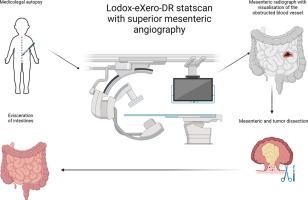
Abstract Image

翻开肠系膜:应用 LODOX eXero-DR® statscan 快速进行死后血管造影。
理由经济实惠的 LODOX eXero-DR® statscan 扫描仪可在资源有限的环境中为法医病理学家提供帮助。使用 LODOX eXero-DR® 进行死后血管造影有助于检测血管病变,从而提高常规尸检的效率。方法我们展示了一例患有滤泡性淋巴瘤的 63 岁男性病例,他在肠系膜淋巴结活检 11 天后出现小肠穿孔。在尸检过程中,切除了肠系膜,露出了肠系膜上动脉源头。使用 Foley 导管将造影剂以 5 毫升为单位注入动脉,然后进行 LODOX eXero-DR® 成像。此过程重复五次,准确定位血管阻塞部位。使用 LODOX eXero-DR® 进行血管造影获得了重要信息,有助于病理鉴定和建立病历。值得注意的是,这是 LODOX eXero-DR® statscan 首次用于尸检血管造影。
本文章由计算机程序翻译,如有差异,请以英文原文为准。
求助全文
约1分钟内获得全文 求助全文
来源期刊
Forensic Imaging
Forensic Imaging RADIOLOGY, NUCLEAR MEDICINE & MEDICAL IMAGING-
CiteScore
2.20
自引率
27.30%
发文量
39
×
引用
GB/T 7714-2015
复制
MLA
复制
APA
复制
导出至
BibTeX EndNote RefMan NoteFirst NoteExpress
×
提示
您的信息不完整,为了账户安全,请先补充。
现在去补充
×
提示
您因"违规操作"
具体请查看互助需知
我知道了
×
提示
确定
请完成安全验证×
copy
已复制链接
快去分享给好友吧!
我知道了
右上角分享
点击右上角分享
0
联系我们:info@booksci.cn Book学术提供免费学术资源搜索服务,方便国内外学者检索中英文文献。致力于提供最便捷和优质的服务体验。 Copyright © 2023 布克学术 All rights reserved.
京ICP备2023020795号-1
ghs 京公网安备 11010802042870号
Book学术文献互助
Book学术文献互助群
群 号:481959085
Book学术官方微信