Matthew Luce DO, Ryan Brandt DO, Joseph Betcher MD
{"title":"Point-of-care ultrasound identifies surgical emergency, expediting care","authors":"Matthew Luce DO, Ryan Brandt DO, Joseph Betcher MD","doi":"10.1002/emp2.13327","DOIUrl":null,"url":null,"abstract":"<p>A 62-year-old male with a history of alcoholic cirrhosis with esophageal varices presented with a chief complaint of hematemesis and abdominal distention. Examination demonstrated ascites and a long-standing umbilical hernia. Given the patient's worsening pain and ongoing hematemesis, point-of-care ultrasound (POCUS) was utilized (Figure 1), which revealed the diagnosis, and was later confirmed with a contrast-enhanced computed tomography (CT) (Figure 2).</p><p>In this case, POCUS was utilized and accurately identified a closed-loop bowel obstruction suspended in the ascites fluid (Video 1). A contrast-enhanced CT confirmed an incarcerated umbilical hernia, and the patient was brought to the operating room for an umbilical hernia repair and small bowel release, as well as gastrointestinal consultation for possible esophageal variceal bleeding.</p><p>The current gold-standard imaging modality for small bowel obstruction (SBO) is CT imaging. This case demonstrates the utility of POCUS in the diagnosis of SBO at bedside (Video 2). Considering his extensive history of high-risk cirrhosis leading to hematemesis and a challenging abdominal examination revealing long-standing ascites, treating physicians may face the risk of anchoring bias, potentially narrowing their focus on the possibility of esophageal variceal bleeding. POCUS quickly revealed the additional pathology, with the obstruction evident within the ascites. POCUS has also demonstrated a significant reduction in time to imaging completion when utilized for bowel obstructions, potentially leading to shorter time to surgical intervention.<span><sup>1</sup></span> Depending on certain clinical factors, some patients may be able to forego CT scans after demonstration of an obstruction process on POCUS.<span><sup>2</sup></span></p><p>All authors contributed significantly to the preparation of this report.</p><p>The authors declare they have no conflicts of interest.</p><p>The authors received no specific funding for this work.</p>","PeriodicalId":73967,"journal":{"name":"Journal of the American College of Emergency Physicians open","volume":"5 6","pages":""},"PeriodicalIF":1.6000,"publicationDate":"2024-11-08","publicationTypes":"Journal Article","fieldsOfStudy":null,"isOpenAccess":false,"openAccessPdf":"https://www.ncbi.nlm.nih.gov/pmc/articles/PMC11549060/pdf/","citationCount":"0","resultStr":null,"platform":"Semanticscholar","paperid":null,"PeriodicalName":"Journal of the American College of Emergency Physicians open","FirstCategoryId":"1085","ListUrlMain":"https://onlinelibrary.wiley.com/doi/10.1002/emp2.13327","RegionNum":0,"RegionCategory":null,"ArticlePicture":[],"TitleCN":null,"AbstractTextCN":null,"PMCID":null,"EPubDate":"","PubModel":"","JCR":"Q2","JCRName":"EMERGENCY MEDICINE","Score":null,"Total":0}
引用次数: 0
Abstract
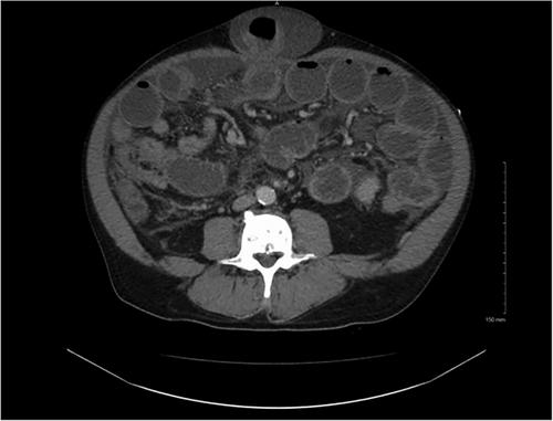
A 62-year-old male with a history of alcoholic cirrhosis with esophageal varices presented with a chief complaint of hematemesis and abdominal distention. Examination demonstrated ascites and a long-standing umbilical hernia. Given the patient's worsening pain and ongoing hematemesis, point-of-care ultrasound (POCUS) was utilized (Figure 1), which revealed the diagnosis, and was later confirmed with a contrast-enhanced computed tomography (CT) (Figure 2).
In this case, POCUS was utilized and accurately identified a closed-loop bowel obstruction suspended in the ascites fluid (Video 1). A contrast-enhanced CT confirmed an incarcerated umbilical hernia, and the patient was brought to the operating room for an umbilical hernia repair and small bowel release, as well as gastrointestinal consultation for possible esophageal variceal bleeding.
The current gold-standard imaging modality for small bowel obstruction (SBO) is CT imaging. This case demonstrates the utility of POCUS in the diagnosis of SBO at bedside (Video 2). Considering his extensive history of high-risk cirrhosis leading to hematemesis and a challenging abdominal examination revealing long-standing ascites, treating physicians may face the risk of anchoring bias, potentially narrowing their focus on the possibility of esophageal variceal bleeding. POCUS quickly revealed the additional pathology, with the obstruction evident within the ascites. POCUS has also demonstrated a significant reduction in time to imaging completion when utilized for bowel obstructions, potentially leading to shorter time to surgical intervention.1 Depending on certain clinical factors, some patients may be able to forego CT scans after demonstration of an obstruction process on POCUS.2
All authors contributed significantly to the preparation of this report.
The authors declare they have no conflicts of interest.
The authors received no specific funding for this work.

 求助内容:
求助内容: 应助结果提醒方式:


