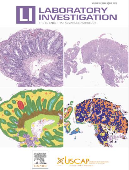
Lymph Node Metastasis Prediction From In Situ Lung Squamous Cell Carcinoma Histopathology Images Using Deep Learning
IF 5.1
2区 医学
Q1 MEDICINE, RESEARCH & EXPERIMENTAL
引用次数: 0
Abstract
Lung squamous cell carcinoma (LUSC), a subtype of non–small cell lung cancer, represents a significant portion of lung cancer cases with distinct histologic patterns impacting prognosis and treatment. The current pathological assessment methods face limitations such as interobserver variability, necessitating more reliable techniques. This study seeks to predict lymph node metastasis in LUSC using deep learning models applied to histopathology images of primary tumors, offering a more accurate and objective method for diagnosis and prognosis. Whole slide images (WSIs) from the Outdo-LUSC and the cancer genome atlas cohorts were used to train and validate deep learning models. Multiinstance learning was applied, with patch-level predictions aggregated into WSI-level outcomes. The study employed the ResNet-18 network, transfer learning, and rigorous data preprocessing. To represent WSI features, innovative techniques like patch likelihood histogram and bag of words were used, followed by training of machine learning classifiers, including the ExtraTrees algorithm. The diagnostic model for lymph node metastasis showed strong performance, particularly using the ExtraTrees algorithm, as demonstrated by receiver operating characteristic curves and gradient-weighted class activation mapping visualizations. The signature generated by the ExtraTrees algorithm, named lymph node status-related in situ LUSC histopathology (LN_ISLUSCH), achieved an area under the curve of 0.941 (95% CI: 0.926-0.955) in the training set and 0.788 (95% CI: 0.748-0.827) in the test set. Kaplan-Meier analyses confirmed that the LN_ISLUSCH model was a significant prognostic factor (P = .02). This study underscores the potential of artificial intelligence in enhancing diagnostic precision in pathology. The LN_ISLUSCH model stands out as a promising tool for predicting lymph node metastasis and prognosis in LUSC. Future studies should focus on larger and more diverse cohorts and explore the integration of additional omics data to further refine predictive accuracy and clinical utility.
利用深度学习从原位肺鳞癌组织病理学图像中预测淋巴结转移。
肺鳞状细胞癌(LUSC)是非小细胞肺癌的一个亚型,在肺癌病例中占很大比例,其独特的组织学模式对预后和治疗产生影响。目前的病理评估方法存在观察者之间的差异等局限性,因此需要更可靠的技术。本研究旨在利用应用于原发性肿瘤组织病理学图像的深度学习模型预测肺癌淋巴结转移,为诊断和预后提供更准确、更客观的方法。来自Outdo-LUSC和TCGA-LUSC队列的全切片图像(WSI)被用于训练和验证深度学习模型。应用多实例学习,将斑块级预测结果汇总为WSI级结果。研究采用了 ResNet-18 网络、迁移学习和严格的数据预处理。为了表示 WSI 特征,使用了斑块似然直方图(PLH)和词包(BoW)等创新技术,然后训练机器学习分类器,包括 ExtraTrees 算法。淋巴结转移诊断模型显示出很强的性能,尤其是使用 ExtraTrees 算法时,接收器操作特征曲线(ROC)和 Grad-CAM 可视化效果都证明了这一点。由 ExtraTrees 算法生成的特征被命名为 LN_ISLUSCH(淋巴结状态相关的原位肺鳞癌组织病理学),在训练集中的曲线下面积(AUC)为 0.941(95% CI:0.926-0.955),在测试集中的曲线下面积(AUC)为 0.788(95% CI:0.748-0.827)。Kaplan-Meier 分析证实,LN_ISLUSCH 模型是一个重要的预后因素(p = 0.02)。这项研究强调了人工智能在提高病理诊断精确度方面的潜力。LN_ISLUSCH 模型是预测 LUSC 淋巴结转移和预后的有效工具。未来的研究应侧重于更大、更多样化的队列,并探索整合更多的全息数据,以进一步提高预测准确性和临床实用性。
本文章由计算机程序翻译,如有差异,请以英文原文为准。
求助全文
约1分钟内获得全文
求助全文
来源期刊

Laboratory Investigation
医学-病理学
CiteScore
8.30
自引率
0.00%
发文量
125
审稿时长
2 months
期刊介绍:
Laboratory Investigation is an international journal owned by the United States and Canadian Academy of Pathology. Laboratory Investigation offers prompt publication of high-quality original research in all biomedical disciplines relating to the understanding of human disease and the application of new methods to the diagnosis of disease. Both human and experimental studies are welcome.
 求助内容:
求助内容: 应助结果提醒方式:


