Application of fluorescent cholangiography to complex biliary variants of the confluence of the cystic duct and the infraportal type of the left lateral bile duct during single-incision laparoscopic cholecystectomy: A case report
{"title":"Application of fluorescent cholangiography to complex biliary variants of the confluence of the cystic duct and the infraportal type of the left lateral bile duct during single-incision laparoscopic cholecystectomy: A case report","authors":"Shinji Nishino, Tsuyoshi Igami, Yukihiro Yokoyama, Takashi Mizuno, Junpei Yamaguchi, Shunsuke Onoe, Masaki Sunagawa, Nobuyuki Watanabe, Taisuke Baba, Shoji Kawakatsu, Tomoki Ebata","doi":"10.1111/ases.13404","DOIUrl":null,"url":null,"abstract":"<p>A 21-year-old man was diagnosed with segmental adenomyomatosis of the gallbladder based on ultrasonography and computed tomography images. Computed tomography with drip infusion cholangiography revealed that the cystic duct joined the infraportal type of the left lateral bile duct (IPLLBD), which runs caudal to the umbilical portion, and that the left medial bile duct joined the right hepatic duct without forming the left hepatic duct. We planned a single-incision laparoscopic cholecystectomy with fluorescent cholangiography. The fluorescent cholangiography visualized the anatomic variant of the biliary system, and the cystic duct was divided safely. Fluorescent cholangiography is a suitable procedure to depict complex biliary anatomic variations in this patient. IPLLBD without the formation of the left hepatic duct is potentially hazardous during cholecystectomy.</p>","PeriodicalId":47019,"journal":{"name":"Asian Journal of Endoscopic Surgery","volume":"18 1","pages":""},"PeriodicalIF":0.9000,"publicationDate":"2024-11-07","publicationTypes":"Journal Article","fieldsOfStudy":null,"isOpenAccess":false,"openAccessPdf":"https://onlinelibrary.wiley.com/doi/epdf/10.1111/ases.13404","citationCount":"0","resultStr":null,"platform":"Semanticscholar","paperid":null,"PeriodicalName":"Asian Journal of Endoscopic Surgery","FirstCategoryId":"1085","ListUrlMain":"https://onlinelibrary.wiley.com/doi/10.1111/ases.13404","RegionNum":0,"RegionCategory":null,"ArticlePicture":[],"TitleCN":null,"AbstractTextCN":null,"PMCID":null,"EPubDate":"","PubModel":"","JCR":"Q4","JCRName":"ORTHOPEDICS","Score":null,"Total":0}
引用次数: 0
Abstract
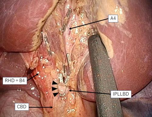
A 21-year-old man was diagnosed with segmental adenomyomatosis of the gallbladder based on ultrasonography and computed tomography images. Computed tomography with drip infusion cholangiography revealed that the cystic duct joined the infraportal type of the left lateral bile duct (IPLLBD), which runs caudal to the umbilical portion, and that the left medial bile duct joined the right hepatic duct without forming the left hepatic duct. We planned a single-incision laparoscopic cholecystectomy with fluorescent cholangiography. The fluorescent cholangiography visualized the anatomic variant of the biliary system, and the cystic duct was divided safely. Fluorescent cholangiography is a suitable procedure to depict complex biliary anatomic variations in this patient. IPLLBD without the formation of the left hepatic duct is potentially hazardous during cholecystectomy.

 求助内容:
求助内容: 应助结果提醒方式:


