Fathy Mohamed Abo Elftouh Elsalhy, Mahmoud Mohammed Ahmed Ali, Mahmoud Fawzy Zaky Morsy, Abdelrahman Ahmed Ali Khattab, Ezzat Nabil Abbas Ibrahim, Hazem Elbadry Mohammed Mohammed, Ramadan Mohamed Abdelrahman Elgohary, Hossam El Din Hassan El Sayed El Baz, Mohamed Sayed Taha Abouzeid
{"title":"Multifocal electroretinogram changes after panretinal photocoagulation in early proliferative diabetic retinopathy.","authors":"Fathy Mohamed Abo Elftouh Elsalhy, Mahmoud Mohammed Ahmed Ali, Mahmoud Fawzy Zaky Morsy, Abdelrahman Ahmed Ali Khattab, Ezzat Nabil Abbas Ibrahim, Hazem Elbadry Mohammed Mohammed, Ramadan Mohamed Abdelrahman Elgohary, Hossam El Din Hassan El Sayed El Baz, Mohamed Sayed Taha Abouzeid","doi":"10.51329/mehdiophthal1503","DOIUrl":null,"url":null,"abstract":"<p><strong>Background: </strong>Panretinal photocoagulation (PRP) impacts macular function in eyes with early proliferative diabetic retinopathy (PDR). Herein, we used the multifocal electroretinogram (mfERG) to objectively investigate this concept.</p><p><strong>Methods: </strong>In this prospective interventional case series, we enrolled patients with treatment-naive early PDR, absence of clinically significant macular edema, and requirement for PRP. All participants underwent detailed ocular examinations. We measured the best-corrected distance visual acuity (BCDVA), conducted optical coherence tomography imaging to measure central macular thickness (CMT), and performed mfERG at baseline and 3 months post-PRP. Amplitude and latency of the mfERG response were evaluated within the innermost four of the five concentric rings.</p><p><strong>Results: </strong>We enrolled 29 eyes of 23 patients with a mean (standard deviation) age of 54.3 (8.8) years and male-to-female ratio of 1:1.3. The mean BCDVA was unchanged post-treatment (<i>P</i> > 0.05), and the BCDVA in 26 eyes (89.7%) was either improved or unchanged, whereas in three eyes (10.3%) it decreased. The mean CMT was unchanged post-PRP (<i>P</i> > 0.05). Concerning the mfERG, the mean P1 amplitudes decreased significantly in all four concentric rings from the foveola at 3 months post-PRP compared with baseline values (all <i>P</i> < 0.05); however, the latencies were unchanged (all <i>P</i> > 0.05). At baseline, BCDVA correlated significantly with both the amplitude (r = + 0.55; <i>P</i> < 0.05) and latency (r = - 0.38; <i>P</i> < 0.05) of the mfERG in the central ring, whereas a significant correlation was detected with only the amplitude at 3 months post-PRP (r = + 0.52; <i>P</i> < 0.05).</p><p><strong>Conclusions: </strong>Macular function was decreased 3 months post-PRP in patients with early PDR, as indicated by decreased amplitude of the mfERG, whereas the functional and anatomical parameters were stable. The mfERG served as an objective tool for measuring retinal function and predicting visual outcomes post-PRP in eyes with early PDR. A higher amplitude in the mfERG correlated substantially with a better visual outcome post-PRP. Further multi-center longitudinal studies with robust designs including different PDR severity levels may reveal additional objective after-effects of PRP<b>.</b></p>","PeriodicalId":36524,"journal":{"name":"Medical Hypothesis, Discovery, and Innovation in Ophthalmology","volume":"13 3","pages":"121-128"},"PeriodicalIF":0.0000,"publicationDate":"2024-10-14","publicationTypes":"Journal Article","fieldsOfStudy":null,"isOpenAccess":false,"openAccessPdf":"https://www.ncbi.nlm.nih.gov/pmc/articles/PMC11537239/pdf/","citationCount":"0","resultStr":null,"platform":"Semanticscholar","paperid":null,"PeriodicalName":"Medical Hypothesis, Discovery, and Innovation in Ophthalmology","FirstCategoryId":"1085","ListUrlMain":"https://doi.org/10.51329/mehdiophthal1503","RegionNum":0,"RegionCategory":null,"ArticlePicture":[],"TitleCN":null,"AbstractTextCN":null,"PMCID":null,"EPubDate":"2024/1/1 0:00:00","PubModel":"eCollection","JCR":"Q2","JCRName":"Medicine","Score":null,"Total":0}
引用次数: 0
Abstract
Background: Panretinal photocoagulation (PRP) impacts macular function in eyes with early proliferative diabetic retinopathy (PDR). Herein, we used the multifocal electroretinogram (mfERG) to objectively investigate this concept.
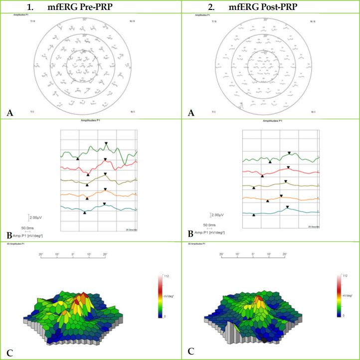
Methods: In this prospective interventional case series, we enrolled patients with treatment-naive early PDR, absence of clinically significant macular edema, and requirement for PRP. All participants underwent detailed ocular examinations. We measured the best-corrected distance visual acuity (BCDVA), conducted optical coherence tomography imaging to measure central macular thickness (CMT), and performed mfERG at baseline and 3 months post-PRP. Amplitude and latency of the mfERG response were evaluated within the innermost four of the five concentric rings.
Results: We enrolled 29 eyes of 23 patients with a mean (standard deviation) age of 54.3 (8.8) years and male-to-female ratio of 1:1.3. The mean BCDVA was unchanged post-treatment (P > 0.05), and the BCDVA in 26 eyes (89.7%) was either improved or unchanged, whereas in three eyes (10.3%) it decreased. The mean CMT was unchanged post-PRP (P > 0.05). Concerning the mfERG, the mean P1 amplitudes decreased significantly in all four concentric rings from the foveola at 3 months post-PRP compared with baseline values (all P < 0.05); however, the latencies were unchanged (all P > 0.05). At baseline, BCDVA correlated significantly with both the amplitude (r = + 0.55; P < 0.05) and latency (r = - 0.38; P < 0.05) of the mfERG in the central ring, whereas a significant correlation was detected with only the amplitude at 3 months post-PRP (r = + 0.52; P < 0.05).
Conclusions: Macular function was decreased 3 months post-PRP in patients with early PDR, as indicated by decreased amplitude of the mfERG, whereas the functional and anatomical parameters were stable. The mfERG served as an objective tool for measuring retinal function and predicting visual outcomes post-PRP in eyes with early PDR. A higher amplitude in the mfERG correlated substantially with a better visual outcome post-PRP. Further multi-center longitudinal studies with robust designs including different PDR severity levels may reveal additional objective after-effects of PRP.

 求助内容:
求助内容: 应助结果提醒方式:


