Kostas G. Mavrakis, Gerasimos Divaris, Maria Tampakaki, Saba N. Khan, Kishan Dholakia, Giannis Zacharakis
{"title":"Optically generated droplet beams improve optoacoustic imaging of choroid thickness as an Alzheimer’s disease biomarker","authors":"Kostas G. Mavrakis, Gerasimos Divaris, Maria Tampakaki, Saba N. Khan, Kishan Dholakia, Giannis Zacharakis","doi":"10.1038/s44310-024-00036-3","DOIUrl":null,"url":null,"abstract":"Optoacoustic microscopy faces a restricted depth of field attributed to the tightly focused Gaussian beam excitation. This limitation poses challenges in capturing high-resolution images of samples with uneven surfaces or obtaining high-quality volumetric images without z-scanning. To address this issue, we propose the use of droplet beam illumination in optoacoustic microscopy, which extends the depth of field to approximately 80 times the Rayleigh length. The droplet beam is generated using a Mach–Zehnder-type interferometer, with each arm equipped with a lens of different optical power. We demonstrate the advantages of droplet beam illumination in microscopy by showing high contrast images on fluorescent beads with a 50% improvement compared to Bessel beam illumination and subsequently imaging the posterior cavity of mice eyes. This method introduces novel perspectives to medical sciences, allowing the measurement of the choroidal layer thickness, an early indicative biomarker for Alzheimer’s disease.","PeriodicalId":501711,"journal":{"name":"npj Nanophotonics","volume":" ","pages":"1-9"},"PeriodicalIF":0.0000,"publicationDate":"2024-11-05","publicationTypes":"Journal Article","fieldsOfStudy":null,"isOpenAccess":false,"openAccessPdf":"https://www.nature.com/articles/s44310-024-00036-3.pdf","citationCount":"0","resultStr":null,"platform":"Semanticscholar","paperid":null,"PeriodicalName":"npj Nanophotonics","FirstCategoryId":"1085","ListUrlMain":"https://www.nature.com/articles/s44310-024-00036-3","RegionNum":0,"RegionCategory":null,"ArticlePicture":[],"TitleCN":null,"AbstractTextCN":null,"PMCID":null,"EPubDate":"","PubModel":"","JCR":"","JCRName":"","Score":null,"Total":0}
引用次数: 0
Abstract
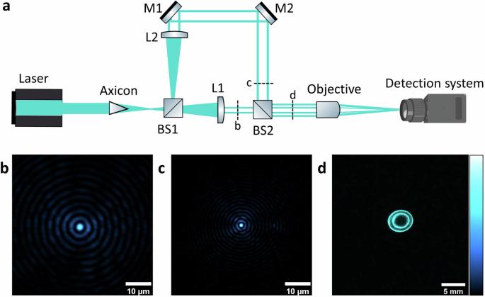
Optoacoustic microscopy faces a restricted depth of field attributed to the tightly focused Gaussian beam excitation. This limitation poses challenges in capturing high-resolution images of samples with uneven surfaces or obtaining high-quality volumetric images without z-scanning. To address this issue, we propose the use of droplet beam illumination in optoacoustic microscopy, which extends the depth of field to approximately 80 times the Rayleigh length. The droplet beam is generated using a Mach–Zehnder-type interferometer, with each arm equipped with a lens of different optical power. We demonstrate the advantages of droplet beam illumination in microscopy by showing high contrast images on fluorescent beads with a 50% improvement compared to Bessel beam illumination and subsequently imaging the posterior cavity of mice eyes. This method introduces novel perspectives to medical sciences, allowing the measurement of the choroidal layer thickness, an early indicative biomarker for Alzheimer’s disease.

 求助内容:
求助内容: 应助结果提醒方式:


