Hao Dun MD MSc , Maura Sticco-Ivins , Yuriko Terada MD PhD , Amber Berning MD , Kory J. Lavine MD PhD , Daniel Kreisel MD PhD , Benjamin J. Kopecky MD PhD
{"title":"Cervical heterotopic heart transplantation in mice using a novel suture technique","authors":"Hao Dun MD MSc , Maura Sticco-Ivins , Yuriko Terada MD PhD , Amber Berning MD , Kory J. Lavine MD PhD , Daniel Kreisel MD PhD , Benjamin J. Kopecky MD PhD","doi":"10.1016/j.jhlto.2024.100164","DOIUrl":null,"url":null,"abstract":"<div><h3>Background</h3><div>Vascularized transplantation models in mice are critical to understand mechanisms that mediate rejection and to develop new therapeutics. Standard abdominal heterotopic heart transplantation techniques employ an <em>end-to-side</em> suture technique and are the workhouse of transplant immunology research laboratories. Recently, cervical heterotopic heart transplantation in mice has emerged as an alternative due to several advantages but is conventionally performed by suture or cuff techniques in an <em>end-to-end</em> fashion. Therefore, we introduce an <em>end-to-side</em> anastomosis technique.</div></div><div><h3>Methods</h3><div>The donor pulmonary artery is <em>end-to-side</em> anastomosed to the recipient right external jugular vein, using a continuous 10–0 nylon suture. Vascular suturing is accomplished inside the vessel on the posterior wall, and then outside the vessel on the anterior wall. Finally, the donor ascending aorta is <em>end-to-side</em> anastomosed to the recipient common carotid artery with an identical suture technique.</div></div><div><h3>Results</h3><div>The median times for the donor heart harvest, recipient preparation, anastomoses of the pulmonary artery to the external jugular vein, and the ascending aorta to the common carotid artery were 12, 10, 12 and 11 minutes, respectively. The survival rate was 100% (<em>n</em> = 20).</div></div><div><h3>Conclusions</h3><div>We provide a detailed description of how to perform <em>end-to-side</em> anastomoses using a suture technique in the mouse cervical heart transplantation model. This procedure reconstitutes coronary blood flow in the heart graft with minimal interruption to recipient anatomy and provides an experimental platform to study transplant immunology.</div></div>","PeriodicalId":100741,"journal":{"name":"JHLT Open","volume":"7 ","pages":"Article 100164"},"PeriodicalIF":0.0000,"publicationDate":"2024-10-17","publicationTypes":"Journal Article","fieldsOfStudy":null,"isOpenAccess":false,"openAccessPdf":"","citationCount":"0","resultStr":null,"platform":"Semanticscholar","paperid":null,"PeriodicalName":"JHLT Open","FirstCategoryId":"1085","ListUrlMain":"https://www.sciencedirect.com/science/article/pii/S2950133424001137","RegionNum":0,"RegionCategory":null,"ArticlePicture":[],"TitleCN":null,"AbstractTextCN":null,"PMCID":null,"EPubDate":"","PubModel":"","JCR":"","JCRName":"","Score":null,"Total":0}
引用次数: 0
Abstract
Background
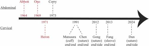
Vascularized transplantation models in mice are critical to understand mechanisms that mediate rejection and to develop new therapeutics. Standard abdominal heterotopic heart transplantation techniques employ an end-to-side suture technique and are the workhouse of transplant immunology research laboratories. Recently, cervical heterotopic heart transplantation in mice has emerged as an alternative due to several advantages but is conventionally performed by suture or cuff techniques in an end-to-end fashion. Therefore, we introduce an end-to-side anastomosis technique.
Methods
The donor pulmonary artery is end-to-side anastomosed to the recipient right external jugular vein, using a continuous 10–0 nylon suture. Vascular suturing is accomplished inside the vessel on the posterior wall, and then outside the vessel on the anterior wall. Finally, the donor ascending aorta is end-to-side anastomosed to the recipient common carotid artery with an identical suture technique.
Results
The median times for the donor heart harvest, recipient preparation, anastomoses of the pulmonary artery to the external jugular vein, and the ascending aorta to the common carotid artery were 12, 10, 12 and 11 minutes, respectively. The survival rate was 100% (n = 20).
Conclusions
We provide a detailed description of how to perform end-to-side anastomoses using a suture technique in the mouse cervical heart transplantation model. This procedure reconstitutes coronary blood flow in the heart graft with minimal interruption to recipient anatomy and provides an experimental platform to study transplant immunology.

 求助内容:
求助内容: 应助结果提醒方式:


