Multinucleated tumor cells and micropapillary morphology appear to be predictors of poor prognosis in renal cell carcinoma with papillary and oncocytic features
IF 2.7
2区 医学
Q2 PATHOLOGY
引用次数: 0
Abstract
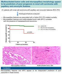
Renal cell carcinoma with papillary and oncocytic features (RCC-PO) are poorly understood, partially due to conflicting results in multiple studies. The histological features that predict behavior of RCC-PO have not been elucidated. The aim is to review clinicopathologic features and to correlate clinical outcomes of patients with RCC-PO to further expand our knowledge on these heterogeneous tumors. An archival search was done for “RCC” and “papillary,” and tumors with >50% papillary and oncocytic features were included. Clinicopathologic data including tumor size, grade, stage, molecular and immunohistochemical testing when performed, and follow-up data were collected. Using multivariate analyses, correlation between histological features, tumor stage and prognosis were analyzed. Sixty-one patients with RCC-PO were identified of which 49 (80%) were male with a median age of 65 (range: 36–93) years, and a mean tumor size of 5.2 (range: 1–21.5) cm. Micropapillary features were seen in 4, bizarre nuclei (at least 3 times larger or with irregular shape) in 6, multinucleated tumor cells (MTC) in 15, single or small clusters (SSC) (made of 2–3 tumor cells) located away from areas of necrosis in 16, and striking eosinophilic cytoplasmic inclusions in 3 tumors, respectively. Thirty-six (59%) tumors were high-grade (WHO/ISUP grade 3–4), and 23 (38%) had a high stage (≥pT3 or pN1). Tumors were positive for AMACR (15/16) and CK7 (13/17), with preserved FH (7/7) staining and were all negative for CD117 (0/7), ALK, TFE3, cathepsin K, Melan A, and HMB45 (0/4, each). Three tumors underwent chromosomal microarray (CMA) plus gene fusion assay, and FISH and germline testing for FLCN and MET gene alterations by PCR were done on 1 each. Ten (16%) patients had a local recurrence (LR) or metastasis after nephrectomy; 4 died of disease (2 had tumors with micropapillary features), with a median follow-up of 7 (range: 0.01–19) years. Tumors with micropapillary features showed significantly higher RCC-PO-related mortality (50% vs. 3.5%, p < 0.001). In multivariable analysis, SSC correlated with a higher stage (HR: 11.95; p = 0.005); micropapillary features (HR: 18.42; p = 0.017) and MTC (HR: 180.22; p = 0.036) with presence of metastasis/LR; and micropapillary features with a higher RCC-PO-related mortality (HR: 60.35; p = 0.036). RCC-PO are cytogenetically heterogeneous with overlapping features of various renal neoplasms. Micropapillary features and MTC appear to be independent predictors of poor outcomes in these tumors.

多核肿瘤细胞和微乳头状形态似乎是具有乳头状和肿瘤细胞特征的肾细胞癌预后不良的预测因素。
人们对具有乳头状和肿瘤细胞特征的肾细胞癌(RCC-PO)知之甚少,部分原因是多项研究的结果相互矛盾。预测RCC-PO行为的组织学特征尚未阐明。本文旨在回顾 RCC-PO 患者的临床病理特征,并对其临床结局进行相关分析,以进一步拓展我们对这些异质性肿瘤的认识。我们对 "RCC "和 "乳头状 "进行了档案检索,并纳入了乳头状和肿瘤细胞特征大于50%的肿瘤。收集的临床病理数据包括肿瘤大小、分级、分期、分子和免疫组化检测(如进行)以及随访数据。通过多变量分析,分析了组织学特征、肿瘤分期和预后之间的相关性。共发现61例RCC-PO患者,其中49例(80%)为男性,中位年龄为65岁(36-93岁),肿瘤平均大小为5.2厘米(1-21.5厘米)。微乳头状特征见于 4 个肿瘤,奇异核(至少增大 3 倍或形状不规则)见于 6 个肿瘤,多核肿瘤细胞(MTC)见于 15 个肿瘤,单个或小团块(SSC)(由 2-3 个肿瘤细胞组成)远离坏死区域见于 16 个肿瘤,显著的嗜酸性细胞质包涵体见于 3 个肿瘤。36例(59%)肿瘤为高级别(WHO/ISUP 3-4级),23例(38%)为高分期(≥pT3或pN1)。肿瘤AMACR(15/16)和CK7(13/17)阳性,FH(7/7)染色保留,CD117(0/7)、ALK、TFE3、cathepsin K、Melan A和HMB45(各0/4)阴性。有 3 例肿瘤接受了染色体微阵列(CMA)加基因融合检测,另有 1 例接受了 FISH 和通过 PCR 进行的 FLCN 和 MET 基因改变的种系检测。10例(16%)患者在肾切除术后出现局部复发(LR)或转移;4例死于疾病(2例肿瘤具有微乳头状特征),中位随访时间为7年(范围:0.01-19年)。具有微乳头状特征的肿瘤显示出更高的 RCC-PO 相关死亡率(50% 对 3.5%,P
本文章由计算机程序翻译,如有差异,请以英文原文为准。
求助全文
约1分钟内获得全文
求助全文
来源期刊

Human pathology
医学-病理学
CiteScore
5.30
自引率
6.10%
发文量
206
审稿时长
21 days
期刊介绍:
Human Pathology is designed to bring information of clinicopathologic significance to human disease to the laboratory and clinical physician. It presents information drawn from morphologic and clinical laboratory studies with direct relevance to the understanding of human diseases. Papers published concern morphologic and clinicopathologic observations, reviews of diseases, analyses of problems in pathology, significant collections of case material and advances in concepts or techniques of value in the analysis and diagnosis of disease. Theoretical and experimental pathology and molecular biology pertinent to human disease are included. This critical journal is well illustrated with exceptional reproductions of photomicrographs and microscopic anatomy.
 求助内容:
求助内容: 应助结果提醒方式:


