Molybdenum interferes with MMPs/TIMPs expression to reduce the receptivity of porcine endometrial epithelial cells

IF 4.7 2区 医学 Q1 BIOCHEMISTRY & MOLECULAR BIOLOGY
Xiao-Ying Gao , Yan Zhang , Wen-Peng Zhao, Er-Jie Tian, Mohammad Mehdi Ommati, Ji-Cang Wang, Hong-Wei Wang, Bian-Hua Zhou
{"title":"Molybdenum interferes with MMPs/TIMPs expression to reduce the receptivity of porcine endometrial epithelial cells","authors":"Xiao-Ying Gao ,&nbsp;Yan Zhang ,&nbsp;Wen-Peng Zhao,&nbsp;Er-Jie Tian,&nbsp;Mohammad Mehdi Ommati,&nbsp;Ji-Cang Wang,&nbsp;Hong-Wei Wang,&nbsp;Bian-Hua Zhou","doi":"10.1016/j.cbi.2024.111304","DOIUrl":null,"url":null,"abstract":"<div><div>To investigate the effect of trace element molybdenum (Mo) on the receptivity of porcine endometrial epithelial cells (PEECs) and evaluate Mo toxicity and its potential molecular mechanisms, Mo-treated PEECs models were established by incubating the cells with various concentrations of medium containing Mo (0, 0.005, 0.020, 0.200, and 5 mmol/L MoNa<sub>2</sub>O<sub>4</sub>·2H<sub>2</sub>O). The results showed that Mo disrupted the morphology and ultrastructure of PEECs, triggered blurred cell edges, cell swelling, cell cycle arrest, and increased apoptosis. At the molecular level, Mo treatment activated the TGF-β1/SMAD2 and PI3K/AKT1 pathways, causing a significant increase in matrix metalloproteinase (MMP)-9 and MMP-2 protein expression. Accompanied by markedly increased tissue inhibitors matrix metalloproteinase (TIMP)-2 and decreased TIMP-1, the balance of MMP2/TIMP-2 and MMP-9/TIMP-1 were disrupted. Ultimately, the receptivity of PEECs was destroyed by excessive Mo, which is revealed by the significant decrease of receptive marker molecules, including leukemia inhibitory factor (LIF), integrins β3 (ITGβ3), heparin-binding epidermal growth factor (HB-EGF), and vascular endothelial growth factor (VEGF). To sum up, the current study demonstrated the potential toxicity of Mo to PEECs, indicating reproductive toxicity at high Mo concentrations and suggesting that the content of Mo should be evaluated as a potential risk factor.</div></div>","PeriodicalId":274,"journal":{"name":"Chemico-Biological Interactions","volume":"405 ","pages":"Article 111304"},"PeriodicalIF":4.7000,"publicationDate":"2024-10-31","publicationTypes":"Journal Article","fieldsOfStudy":null,"isOpenAccess":false,"openAccessPdf":"","citationCount":"0","resultStr":null,"platform":"Semanticscholar","paperid":null,"PeriodicalName":"Chemico-Biological Interactions","FirstCategoryId":"3","ListUrlMain":"https://www.sciencedirect.com/science/article/pii/S0009279724004502","RegionNum":2,"RegionCategory":"医学","ArticlePicture":[],"TitleCN":null,"AbstractTextCN":null,"PMCID":null,"EPubDate":"","PubModel":"","JCR":"Q1","JCRName":"BIOCHEMISTRY & MOLECULAR BIOLOGY","Score":null,"Total":0}
引用次数: 0

Abstract

To investigate the effect of trace element molybdenum (Mo) on the receptivity of porcine endometrial epithelial cells (PEECs) and evaluate Mo toxicity and its potential molecular mechanisms, Mo-treated PEECs models were established by incubating the cells with various concentrations of medium containing Mo (0, 0.005, 0.020, 0.200, and 5 mmol/L MoNa2O4·2H2O). The results showed that Mo disrupted the morphology and ultrastructure of PEECs, triggered blurred cell edges, cell swelling, cell cycle arrest, and increased apoptosis. At the molecular level, Mo treatment activated the TGF-β1/SMAD2 and PI3K/AKT1 pathways, causing a significant increase in matrix metalloproteinase (MMP)-9 and MMP-2 protein expression. Accompanied by markedly increased tissue inhibitors matrix metalloproteinase (TIMP)-2 and decreased TIMP-1, the balance of MMP2/TIMP-2 and MMP-9/TIMP-1 were disrupted. Ultimately, the receptivity of PEECs was destroyed by excessive Mo, which is revealed by the significant decrease of receptive marker molecules, including leukemia inhibitory factor (LIF), integrins β3 (ITGβ3), heparin-binding epidermal growth factor (HB-EGF), and vascular endothelial growth factor (VEGF). To sum up, the current study demonstrated the potential toxicity of Mo to PEECs, indicating reproductive toxicity at high Mo concentrations and suggesting that the content of Mo should be evaluated as a potential risk factor.
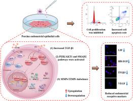
Abstract Image

钼可干扰 MMPs/TIMPs 的表达,从而降低猪子宫内膜上皮细胞的接受能力。
为了研究微量元素钼(Mo)对猪子宫内膜上皮细胞(PEECs)接受性的影响,并评估钼的毒性及其潜在的分子机制,研究人员用不同浓度的含钼培养基(0、0.005、0.020、0.200和5mmol/L MoNa2O4-2H2O)培养猪子宫内膜上皮细胞,建立了钼处理的PEECs模型。结果表明,Mo 破坏了 PEECs 的形态和超微结构,导致细胞边缘模糊、细胞肿胀、细胞周期停滞和细胞凋亡增加。在分子水平上,Mo 处理激活了 TGF-β1/SMAD2 和 PI3K/AKT1 通路,导致基质金属蛋白酶(MMP)-9 和 MMP-2 蛋白表达显著增加。伴随着组织抑制基质金属蛋白酶(TIMP)-2 的明显增加和 TIMP-1 的减少,MMP2/TIMP-2 和 MMP-9/TIMP-1 的平衡被打破。最终,白血病抑制因子(LIF)、整合素β3(ITGβ3)、肝素结合表皮生长因子(HB-EGF)和血管内皮生长因子(VEGF)等接受性标志物分子显著减少,表明过量的 Mo 破坏了 PEECs 的接受性。总之,目前的研究证明了钼对 PEECs 的潜在毒性,表明高浓度钼具有生殖毒性,并建议将钼含量作为潜在风险因素进行评估。
本文章由计算机程序翻译,如有差异,请以英文原文为准。
求助全文
约1分钟内获得全文 求助全文
来源期刊
CiteScore
7.70
自引率
3.90%
发文量
410
审稿时长
36 days
期刊介绍: Chemico-Biological Interactions publishes research reports and review articles that examine the molecular, cellular, and/or biochemical basis of toxicologically relevant outcomes. Special emphasis is placed on toxicological mechanisms associated with interactions between chemicals and biological systems. Outcomes may include all traditional endpoints caused by synthetic or naturally occurring chemicals, both in vivo and in vitro. Endpoints of interest include, but are not limited to carcinogenesis, mutagenesis, respiratory toxicology, neurotoxicology, reproductive and developmental toxicology, and immunotoxicology.
×
引用
GB/T 7714-2015
复制
MLA
复制
APA
复制
导出至
BibTeX EndNote RefMan NoteFirst NoteExpress
×
提示
您的信息不完整,为了账户安全,请先补充。
现在去补充
×
提示
您因"违规操作"
具体请查看互助需知
我知道了
×
提示
确定
请完成安全验证×
copy
已复制链接
快去分享给好友吧!
我知道了
右上角分享
点击右上角分享
0
联系我们:info@booksci.cn Book学术提供免费学术资源搜索服务,方便国内外学者检索中英文文献。致力于提供最便捷和优质的服务体验。 Copyright © 2023 布克学术 All rights reserved.
京ICP备2023020795号-1
ghs 京公网安备 11010802042870号
Book学术文献互助
Book学术文献互助群
群 号:481959085
Book学术官方微信