The impact of cathepsins on liver hepatocellular carcinoma: Insights from genetic and functional analyses

IF 2.6 3区 生物学 Q2 GENETICS & HEREDITY
Gene Pub Date : 2024-10-30 DOI:10.1016/j.gene.2024.149064
Qi Liu , Junyi Chen , Yuyang Liu , Shengwei Zhang , Hui Feng , Tao Wan , Shemin Zhang , Ning Zhang , Zhanyu Yang
{"title":"The impact of cathepsins on liver hepatocellular carcinoma: Insights from genetic and functional analyses","authors":"Qi Liu ,&nbsp;Junyi Chen ,&nbsp;Yuyang Liu ,&nbsp;Shengwei Zhang ,&nbsp;Hui Feng ,&nbsp;Tao Wan ,&nbsp;Shemin Zhang ,&nbsp;Ning Zhang ,&nbsp;Zhanyu Yang","doi":"10.1016/j.gene.2024.149064","DOIUrl":null,"url":null,"abstract":"<div><div>Liver Hepatocellular Carcinoma (LIHC), ranked as the second deadliest cancer globally, poses a major health challenge because of its widespread occurrence and poor prognosis. The mechanisms underlying LIHC development and progression remain unclear. Cathepsins are linked to tumorigenesis in other cancers, but their role in LIHC is underexplored. This study employed integrative analyses, including Mendelian Randomization (MR), bulk RNA-sequencing (bulk-seq), single-cell RNA sequencing (scRNA-seq), immunohistochemical (IHC) analysis, and cellular experiments with siRNA technology, to investigate the role of cathepsin E (CTSE) in LIHC. MR analysis identified CTSE as a factor associated with increased LIHC risk. Prognostic analysis using TCGA data showed that higher CTSE levels are linked to poorer survival, establishing CTSE as an independent prognostic risk factor. Integrative transcriptome analysis revealed close relation of CTSE to the extracellular matrix. scRNA-seq from TISCH2 demonstrated that CTSE is predominantly expressed in malignant LIHC cells. IHC confirmed higher CTSE expression in LIHC tissues compared to peritumoral tissues. Functional assays, such as qRT-PCR, Western blot, cell proliferation, and colony formation experiments, demonstrated that siRNA-mediated CTSE knockdown in HepG2 and Huh7 cell lines notably suppressed cell proliferation and altered the FAK/Paxillin/Akt signaling cascade. This research enhances our comprehension of LIHC development, emphasizing CTSE as a promising prognostic marker and potential therapeutic target. Inhibiting CTSE could slow the progression of LIHC, presenting novel opportunities for therapeutic approaches.</div></div>","PeriodicalId":12499,"journal":{"name":"Gene","volume":"935 ","pages":"Article 149064"},"PeriodicalIF":2.6000,"publicationDate":"2024-10-30","publicationTypes":"Journal Article","fieldsOfStudy":null,"isOpenAccess":false,"openAccessPdf":"","citationCount":"0","resultStr":null,"platform":"Semanticscholar","paperid":null,"PeriodicalName":"Gene","FirstCategoryId":"99","ListUrlMain":"https://www.sciencedirect.com/science/article/pii/S0378111924009454","RegionNum":3,"RegionCategory":"生物学","ArticlePicture":[],"TitleCN":null,"AbstractTextCN":null,"PMCID":null,"EPubDate":"","PubModel":"","JCR":"Q2","JCRName":"GENETICS & HEREDITY","Score":null,"Total":0}
引用次数: 0

Abstract

Liver Hepatocellular Carcinoma (LIHC), ranked as the second deadliest cancer globally, poses a major health challenge because of its widespread occurrence and poor prognosis. The mechanisms underlying LIHC development and progression remain unclear. Cathepsins are linked to tumorigenesis in other cancers, but their role in LIHC is underexplored. This study employed integrative analyses, including Mendelian Randomization (MR), bulk RNA-sequencing (bulk-seq), single-cell RNA sequencing (scRNA-seq), immunohistochemical (IHC) analysis, and cellular experiments with siRNA technology, to investigate the role of cathepsin E (CTSE) in LIHC. MR analysis identified CTSE as a factor associated with increased LIHC risk. Prognostic analysis using TCGA data showed that higher CTSE levels are linked to poorer survival, establishing CTSE as an independent prognostic risk factor. Integrative transcriptome analysis revealed close relation of CTSE to the extracellular matrix. scRNA-seq from TISCH2 demonstrated that CTSE is predominantly expressed in malignant LIHC cells. IHC confirmed higher CTSE expression in LIHC tissues compared to peritumoral tissues. Functional assays, such as qRT-PCR, Western blot, cell proliferation, and colony formation experiments, demonstrated that siRNA-mediated CTSE knockdown in HepG2 and Huh7 cell lines notably suppressed cell proliferation and altered the FAK/Paxillin/Akt signaling cascade. This research enhances our comprehension of LIHC development, emphasizing CTSE as a promising prognostic marker and potential therapeutic target. Inhibiting CTSE could slow the progression of LIHC, presenting novel opportunities for therapeutic approaches.
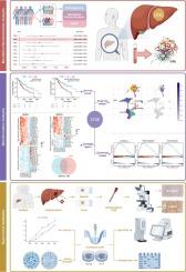
Abstract Image

cathepsins 对肝脏肝细胞癌的影响:遗传和功能分析的启示。
肝细胞癌(LIHC)被列为全球第二大致命癌症,因其发病率高、预后差而对健康构成重大挑战。肝癌细胞癌的发生和发展机制尚不清楚。胰蛋白酶与其他癌症的肿瘤发生有关,但它们在 LIHC 中的作用尚未得到充分探索。本研究采用综合分析方法,包括孟德尔随机化(Mendelian Randomization,MR)、批量RNA测序(bulk-seq)、单细胞RNA测序(scRNA-seq)、免疫组化(IHC)分析和使用siRNA技术的细胞实验,研究鲶鱼蛋白酶E(CTSE)在LIHC中的作用。磁共振分析发现,CTSE是增加LIHC风险的相关因素。利用TCGA数据进行的预后分析表明,较高的CTSE水平与较差的生存率有关,从而确定CTSE是一个独立的预后风险因素。整合转录组分析显示,CTSE与细胞外基质密切相关。来自TISCH2的scRNA-seq表明,CTSE主要在恶性LIHC细胞中表达。IHC 证实,与瘤周组织相比,CTSE 在 LIHC 组织中的表达更高。qRT-PCR、Western印迹、细胞增殖和集落形成实验等功能测试表明,在HepG2和Huh7细胞系中,siRNA介导的CTSE敲除明显抑制了细胞增殖,并改变了FAK/Paxillin/Akt信号级联。这项研究加深了我们对LIHC发展的理解,强调了CTSE是一种有前景的预后标志物和潜在的治疗靶点。抑制CTSE可减缓LIHC的进展,为治疗方法提供新的机遇。
本文章由计算机程序翻译,如有差异,请以英文原文为准。
求助全文
约1分钟内获得全文 求助全文
来源期刊
Gene
Gene 生物-遗传学
CiteScore
6.10
自引率
2.90%
发文量
718
审稿时长
42 days
期刊介绍: Gene publishes papers that focus on the regulation, expression, function and evolution of genes in all biological contexts, including all prokaryotic and eukaryotic organisms, as well as viruses.
×
引用
GB/T 7714-2015
复制
MLA
复制
APA
复制
导出至
BibTeX EndNote RefMan NoteFirst NoteExpress
×
提示
您的信息不完整,为了账户安全,请先补充。
现在去补充
×
提示
您因"违规操作"
具体请查看互助需知
我知道了
×
提示
确定
请完成安全验证×
copy
已复制链接
快去分享给好友吧!
我知道了
右上角分享
点击右上角分享
0
联系我们:info@booksci.cn Book学术提供免费学术资源搜索服务,方便国内外学者检索中英文文献。致力于提供最便捷和优质的服务体验。 Copyright © 2023 布克学术 All rights reserved.
京ICP备2023020795号-1
ghs 京公网安备 11010802042870号
Book学术文献互助
Book学术文献互助群
群 号:481959085
Book学术官方微信