{"title":"Nanoparticle-encapsuled celastrol-Fe(III): “Single-edged sword” for tumor therapy featuring microenvironment-dependent toxicity","authors":"Yu Liu, Zhiyong Qian","doi":"10.1002/mog2.70001","DOIUrl":null,"url":null,"abstract":"<p>Recently, Li et al. published a study in <i>Science Advances</i> on the use of dual-responsive celastrol (CEL) materials for cancer treatment. The study took advantage of the high adenosine triphosphate (ATP) levels in the tumor microenvironment to modify the interior and surface of CEL, reducing its toxicity to normal tissues and maximizing the efficacy of its cancer treatment which contributed significantly to drug delivery for tumor therapy.<span><sup>1</sup></span></p><p>CEL is a drug with anticancer activity isolated from traditional Chinese medicinal herbs.<span><sup>2</sup></span> It inhibits cancer cell growth and survival signaling by binding to Cdc37 and Hsp90 reaction sites, causing disruption of the Hsp90-Cdc37 binding and folding of some of the client proteins.<span><sup>3</sup></span> However, its water stability, bioavailability, and targeting are undesirable, which limited its clinical application. There are currently two main approaches to promote CEL's therapeutic potential in clinic: 1. synthesizing derivatives by surface modification which mainly focuses on C-20 carboxylic acid functionality, alteration at the A ring, and C-6-modification at the B ring, and 2. adopting nanomedicine delivery systems, where liposomes, polymeric micelles, and nanoparticles (NPs) are commonly used. The second approach, however, requires strictly selected materials. Liposomes, with its ease of aggregation and instability and leakage when packaging drugs, and polymeric micelles, with potential toxicity, unclear drug delivery mechanisms, and poor quality/specification control, demand extra considerations in application. In this study an ATP/ROS dual-responsive CEL derivative was designed for tumor therapy by mainly taking advantage of the environment in which ATP levels inside and outside tumor cells are 10<sup>3</sup> to 10<sup>4</sup> times higher than that around normal tissues.<span><sup>4</sup></span> Surface modification of CEL using metal chelation was performed to address the toxicity to normal tissues, and NPs were selected for packaging to promote the release of CEL when it reaches the tumor site. Compared with liposomes and polymeric micelles, CEL-Fe with ATP/ROS dual-response has a well-defined structural study, low drug toxicity, high stability, and ultimately the ability to respond to the specificity of the tumor microenvironment for effective drug release and treatment which improves the biosafety and biocompatibility of the therapeutic drugs. Fe(III) was chosen to coordinately bond with oxygens on C-2 and C-3 in CEL to produce the less toxic CEL-Fe, which is then coated with polymer membrane consisting of thioketal-containing polyethylene glycol-grafted polymer and polyethylene enamine-modified F127 polymer to elevate its permeability and retention effects.<span><sup>5</sup></span> The high concentration of ATP in tumor tissues disrupts the coordination of Fe(III), restoring the pharmacological toxicity of CEL. In response to high level of ROS at tumor site, thioketal groups are degraded to facilitate drug release, and the oncology treatment is therefore initiated (Figure 1A).</p><p>The cell viability of cancer cells and normal cells were tested after treated with different groups: CEL, CEL-Fe, and CEL-Fe-ATP. The results verified the biosafety of CEL-Fe(III) chelate by showing its ATP-dependent cytotoxicity (Figures 1B,C). The mechanism of this property shift between CEL and CEL-Fe was explored by RNA sequencing analysis of A549 cells. PCA gene expression analysis showed that the CEL group and CEL-Fe+ATP group were clustered together, while separated from the CEL+Fe group. Moreover, the calculation of gene expression fold change and <i>p</i> Value plots revealed no significant gene expression difference between the CEL and CEL-Fe+ATP groups, while that of the CEL+Fe group varied. All above indicated the cytotoxicity weakening of the Fe(III) chelation and its restoration by ATP. The Kyoto Encyclopedia of Genes and Genomes (KEGG) analysis and Gene Set Enrichment Analysis showed a high enrichment of ER's protein processing, tumor necrosis factor signaling, and P53 signaling in CEL and CEL-Fe-ATP treatments, and a large degree of overlap of these pathways in CEL-Fe. This demonstrated that the functioning of CEL was related to the “life cycle” of protein, which was consistent with the mechanism of CEL antitumor therapy in the existing studies. The toxicity mechanism of CEL was further investigated where Hsp90/Cdc37 was chosen as the model for detection. Molecular docking and MD simulation revealed that CEL destroys the hydrogen bonds inside Hsp90-Cdc37 by occupying hydrophobic sites to cause protein degradation, while the number of hydrogen bonds destroyed by CEL-Fe is significantly smaller than that of CEL, which reduces its toxicity to the protein complex and cells. The data above provides a basis for a potential effective cancer therapy with this CEL derivative (Figures 1D,E).</p><p>In-vivo biosafety was examined by hema-toxylin and eosin (H&E) staining of tissue sections. Results showed that CEL and CEL NPs were significantly toxic to several vital tissues and organs, which did not occur in the CEL-Fe group. This demonstrated successful discrimination against tumor from normal tissues and organs brought by the metal chelation. Moreover, fluorescence imaging showed strong presence of CEL NPs at tumor sites in both cell-derived xenograft (CDX) and patient-derived xenograft (PDX) tumor models, demonstrating their superior biodispersibility and feasibility in future clinical trials.</p><p>Further studies were conducted by designing additional contrast groups of phosphate-buffered saline, CEL-Fe NPs, and NPs-treated mice with both CDX and PDX models to assess their antitumor ability. After the prescribed cycle, mice treated by CEL-Fe NPs in both models had the smallest tumor volume and best tumor treatment effect. Both H&E staining and TUNEL assay showed that the CEL-Fe NPs group ranked top in terms of tumor necrosis.</p><p>In conclusion, this study successfully synthesized a nanomaterial with ATP/ROS dual-responsiveness targeting mainly the high ATP level of the tumor microenvironment. The external packaging material was destroyed by Fenton reaction in the high ATP/ROS environment, exposing the loaded therapeutic drug CEL-Fe which was converted into CEL with toxicity restored by the high ATP concentration, exerting its full effect at the tumor site. In this study, the CEL modification cleverly solves CEL's therapeutic problem of discrimination between normal tissues and cancer tissues to achieve the ultimate goal of precision therapy at the tumor sites. This type of design is also the one of the main ideas of current tumor therapy, which takes advantage of the differences between the tumor microenvironment and that of the normal tissues. Such differences include ATP levels, pH, oxygen content, and so forthetc. It is highly expected that researchers may make good use of the intrinsic properties of tumor microenvironment to continue to study and develop drugs or drug delivery systems with ideal effectiveness, efficiency, and targeting.</p><p><b>Yu Liu</b>: Conception; drafting of the manuscript. <b>Zhiyong Qian</b>: Supervision. Both authors have read and approved the final version of the article.</p><p>The authors declare no conflict of interest.</p><p>Not applicable.</p>","PeriodicalId":100902,"journal":{"name":"MedComm – Oncology","volume":"3 4","pages":""},"PeriodicalIF":0.0000,"publicationDate":"2024-10-28","publicationTypes":"Journal Article","fieldsOfStudy":null,"isOpenAccess":false,"openAccessPdf":"https://onlinelibrary.wiley.com/doi/epdf/10.1002/mog2.70001","citationCount":"0","resultStr":null,"platform":"Semanticscholar","paperid":null,"PeriodicalName":"MedComm – Oncology","FirstCategoryId":"1085","ListUrlMain":"https://onlinelibrary.wiley.com/doi/10.1002/mog2.70001","RegionNum":0,"RegionCategory":null,"ArticlePicture":[],"TitleCN":null,"AbstractTextCN":null,"PMCID":null,"EPubDate":"","PubModel":"","JCR":"","JCRName":"","Score":null,"Total":0}
引用次数: 0
Abstract
Recently, Li et al. published a study in Science Advances on the use of dual-responsive celastrol (CEL) materials for cancer treatment. The study took advantage of the high adenosine triphosphate (ATP) levels in the tumor microenvironment to modify the interior and surface of CEL, reducing its toxicity to normal tissues and maximizing the efficacy of its cancer treatment which contributed significantly to drug delivery for tumor therapy.1
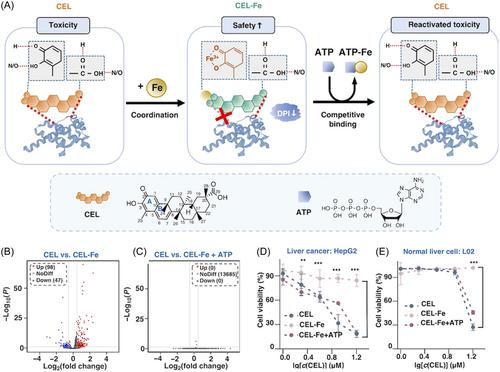
CEL is a drug with anticancer activity isolated from traditional Chinese medicinal herbs.2 It inhibits cancer cell growth and survival signaling by binding to Cdc37 and Hsp90 reaction sites, causing disruption of the Hsp90-Cdc37 binding and folding of some of the client proteins.3 However, its water stability, bioavailability, and targeting are undesirable, which limited its clinical application. There are currently two main approaches to promote CEL's therapeutic potential in clinic: 1. synthesizing derivatives by surface modification which mainly focuses on C-20 carboxylic acid functionality, alteration at the A ring, and C-6-modification at the B ring, and 2. adopting nanomedicine delivery systems, where liposomes, polymeric micelles, and nanoparticles (NPs) are commonly used. The second approach, however, requires strictly selected materials. Liposomes, with its ease of aggregation and instability and leakage when packaging drugs, and polymeric micelles, with potential toxicity, unclear drug delivery mechanisms, and poor quality/specification control, demand extra considerations in application. In this study an ATP/ROS dual-responsive CEL derivative was designed for tumor therapy by mainly taking advantage of the environment in which ATP levels inside and outside tumor cells are 103 to 104 times higher than that around normal tissues.4 Surface modification of CEL using metal chelation was performed to address the toxicity to normal tissues, and NPs were selected for packaging to promote the release of CEL when it reaches the tumor site. Compared with liposomes and polymeric micelles, CEL-Fe with ATP/ROS dual-response has a well-defined structural study, low drug toxicity, high stability, and ultimately the ability to respond to the specificity of the tumor microenvironment for effective drug release and treatment which improves the biosafety and biocompatibility of the therapeutic drugs. Fe(III) was chosen to coordinately bond with oxygens on C-2 and C-3 in CEL to produce the less toxic CEL-Fe, which is then coated with polymer membrane consisting of thioketal-containing polyethylene glycol-grafted polymer and polyethylene enamine-modified F127 polymer to elevate its permeability and retention effects.5 The high concentration of ATP in tumor tissues disrupts the coordination of Fe(III), restoring the pharmacological toxicity of CEL. In response to high level of ROS at tumor site, thioketal groups are degraded to facilitate drug release, and the oncology treatment is therefore initiated (Figure 1A).
The cell viability of cancer cells and normal cells were tested after treated with different groups: CEL, CEL-Fe, and CEL-Fe-ATP. The results verified the biosafety of CEL-Fe(III) chelate by showing its ATP-dependent cytotoxicity (Figures 1B,C). The mechanism of this property shift between CEL and CEL-Fe was explored by RNA sequencing analysis of A549 cells. PCA gene expression analysis showed that the CEL group and CEL-Fe+ATP group were clustered together, while separated from the CEL+Fe group. Moreover, the calculation of gene expression fold change and p Value plots revealed no significant gene expression difference between the CEL and CEL-Fe+ATP groups, while that of the CEL+Fe group varied. All above indicated the cytotoxicity weakening of the Fe(III) chelation and its restoration by ATP. The Kyoto Encyclopedia of Genes and Genomes (KEGG) analysis and Gene Set Enrichment Analysis showed a high enrichment of ER's protein processing, tumor necrosis factor signaling, and P53 signaling in CEL and CEL-Fe-ATP treatments, and a large degree of overlap of these pathways in CEL-Fe. This demonstrated that the functioning of CEL was related to the “life cycle” of protein, which was consistent with the mechanism of CEL antitumor therapy in the existing studies. The toxicity mechanism of CEL was further investigated where Hsp90/Cdc37 was chosen as the model for detection. Molecular docking and MD simulation revealed that CEL destroys the hydrogen bonds inside Hsp90-Cdc37 by occupying hydrophobic sites to cause protein degradation, while the number of hydrogen bonds destroyed by CEL-Fe is significantly smaller than that of CEL, which reduces its toxicity to the protein complex and cells. The data above provides a basis for a potential effective cancer therapy with this CEL derivative (Figures 1D,E).
In-vivo biosafety was examined by hema-toxylin and eosin (H&E) staining of tissue sections. Results showed that CEL and CEL NPs were significantly toxic to several vital tissues and organs, which did not occur in the CEL-Fe group. This demonstrated successful discrimination against tumor from normal tissues and organs brought by the metal chelation. Moreover, fluorescence imaging showed strong presence of CEL NPs at tumor sites in both cell-derived xenograft (CDX) and patient-derived xenograft (PDX) tumor models, demonstrating their superior biodispersibility and feasibility in future clinical trials.
Further studies were conducted by designing additional contrast groups of phosphate-buffered saline, CEL-Fe NPs, and NPs-treated mice with both CDX and PDX models to assess their antitumor ability. After the prescribed cycle, mice treated by CEL-Fe NPs in both models had the smallest tumor volume and best tumor treatment effect. Both H&E staining and TUNEL assay showed that the CEL-Fe NPs group ranked top in terms of tumor necrosis.
In conclusion, this study successfully synthesized a nanomaterial with ATP/ROS dual-responsiveness targeting mainly the high ATP level of the tumor microenvironment. The external packaging material was destroyed by Fenton reaction in the high ATP/ROS environment, exposing the loaded therapeutic drug CEL-Fe which was converted into CEL with toxicity restored by the high ATP concentration, exerting its full effect at the tumor site. In this study, the CEL modification cleverly solves CEL's therapeutic problem of discrimination between normal tissues and cancer tissues to achieve the ultimate goal of precision therapy at the tumor sites. This type of design is also the one of the main ideas of current tumor therapy, which takes advantage of the differences between the tumor microenvironment and that of the normal tissues. Such differences include ATP levels, pH, oxygen content, and so forthetc. It is highly expected that researchers may make good use of the intrinsic properties of tumor microenvironment to continue to study and develop drugs or drug delivery systems with ideal effectiveness, efficiency, and targeting.
Yu Liu: Conception; drafting of the manuscript. Zhiyong Qian: Supervision. Both authors have read and approved the final version of the article.

 求助内容:
求助内容: 应助结果提醒方式:


