Deep neural network model for diagnosing diabetic retinopathy detection: An efficient mechanism for diabetic management
IF 4.9
2区 医学
Q1 ENGINEERING, BIOMEDICAL
引用次数: 0
Abstract
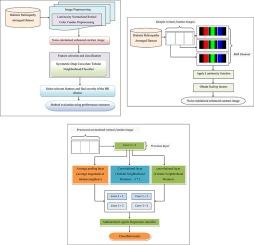
Diabetic retinopathy (DR) is a common eye disease and a notable starting point of blindness in diabetic patients. Detecting the existence of a microaneurysm in the fundus images and the identification of DR in the preliminary stage has been a considerable question for decades. Systematic screening and interference are the most efficient mechanisms for disease management. The sizeable populations of diabetic patients and their enormous screening requirements have given rise to the computer-aided and automatic diagnosis of DR. The utilization of Deep Neural Networks in DR diagnosis has also attracted much attention and considerable advancement has been made. Diabetic retinopathy (DR) includes sensitivity and specificity that are particular to the Diabetic tested (see section on probabilistic reasoning). Even if a test is performed correctly, there is a chance for a false positive or false negative result. However, despite the several advancements that have been made, there remains room for improvement in the sensitivity and specificity of the DR diagnosis. In this work, a novel method called the Luminosity Normalized Symmetric Deep Convolute Tubular Classifier (LN-SDCTC) for DR detection is proposed. The LN-SDCTC method is split into two parts. Initially, with the retinal color fundus images as input, the Luminosity Normalized Retinal Color Fundus Preprocessing model applies to produce a noise-minimized enhanced contrast image. Second, we provide the get-processed image as input to the Symmetric Deep Convolute network. Here, with the aid of the convolutional layer (i.e., the Tubular Neighborhood Window), the average pooling layer (i.e., average magnitude value of tubular neighbors), and the max-pooling layer (i.e., maximum contrast orientation), relevant features are selected. Finally, with the extracted features as input and with the aid of the Multinomial Regression Classification function, the severity of the DR disease is determined. Extensive experimental results in terms of peak signal-to-noise ratio, disease detection time, sensitivity, and specificity reveal that the proposed method of DR detection greatly facilitates the deep learning model and yields better results than various state-of-art methods.

用于诊断糖尿病视网膜病变检测的深度神经网络模型:糖尿病管理的高效机制
糖尿病视网膜病变(DR)是一种常见的眼病,也是糖尿病患者失明的显著起因。几十年来,在眼底图像中检测微动脉瘤的存在以及在初期阶段识别糖尿病视网膜病变一直是一个相当大的问题。系统筛查和干扰是最有效的疾病管理机制。庞大的糖尿病患者群体及其巨大的筛查需求催生了 DR 的计算机辅助自动诊断。深度神经网络在糖尿病视网膜病变诊断中的应用也备受关注,并取得了长足的进步。糖尿病视网膜病变(DR)包括敏感性和特异性,而敏感性和特异性是测试的糖尿病患者所特有的(见概率推理部分)。即使检测结果正确,也有可能出现假阳性或假阴性结果。然而,尽管已经取得了一些进步,但 DR 诊断的灵敏度和特异性仍有待提高。本研究提出了一种用于 DR 检测的新方法,即光度归一化对称深度卷积管状分类器(LN-SDCTC)。LN-SDCTC 方法分为两部分。首先,以视网膜彩色眼底图像为输入,应用亮度归一化视网膜彩色眼底预处理模型生成噪声最小化的增强对比度图像。其次,我们将处理后的图像作为对称深度卷积网络的输入。在此,借助卷积层(即管状邻域窗口)、平均池化层(即管状邻域的平均幅度值)和最大池化层(即最大对比度方向),选择相关特征。最后,将提取的特征作为输入,借助多项式回归分类功能,确定 DR 疾病的严重程度。从峰值信噪比、疾病检测时间、灵敏度和特异性等方面的大量实验结果表明,所提出的 DR 检测方法极大地促进了深度学习模型的发展,并产生了比各种先进方法更好的结果。
本文章由计算机程序翻译,如有差异,请以英文原文为准。
求助全文
约1分钟内获得全文
求助全文
来源期刊

Biomedical Signal Processing and Control
工程技术-工程:生物医学
CiteScore
9.80
自引率
13.70%
发文量
822
审稿时长
4 months
期刊介绍:
Biomedical Signal Processing and Control aims to provide a cross-disciplinary international forum for the interchange of information on research in the measurement and analysis of signals and images in clinical medicine and the biological sciences. Emphasis is placed on contributions dealing with the practical, applications-led research on the use of methods and devices in clinical diagnosis, patient monitoring and management.
Biomedical Signal Processing and Control reflects the main areas in which these methods are being used and developed at the interface of both engineering and clinical science. The scope of the journal is defined to include relevant review papers, technical notes, short communications and letters. Tutorial papers and special issues will also be published.
 求助内容:
求助内容: 应助结果提醒方式:


