IHH–GLI-1–HIF-2α signalling influences hypertrophic chondrocytes to exacerbate osteoarthritis progression

IF 5.9 1区 医学 Q1 ORTHOPEDICS
Chengming Zhang , Ruipeng Zhao , Zhengquan Dong , Yang Liu , Mengrou Liu , Haoqian Li , Yukun Yin , Xianda Che , Gaige Wu , li Guo , Pengcui Li , Xiaochun Wei , Ziquan Yang
{"title":"IHH–GLI-1–HIF-2α signalling influences hypertrophic chondrocytes to exacerbate osteoarthritis progression","authors":"Chengming Zhang ,&nbsp;Ruipeng Zhao ,&nbsp;Zhengquan Dong ,&nbsp;Yang Liu ,&nbsp;Mengrou Liu ,&nbsp;Haoqian Li ,&nbsp;Yukun Yin ,&nbsp;Xianda Che ,&nbsp;Gaige Wu ,&nbsp;li Guo ,&nbsp;Pengcui Li ,&nbsp;Xiaochun Wei ,&nbsp;Ziquan Yang","doi":"10.1016/j.jot.2024.09.008","DOIUrl":null,"url":null,"abstract":"<div><h3>Background</h3><div>Chondrocyte hypertrophy is a potential target for osteoarthritis (OA) treatment, with Indian hedgehog (IHH), glioma-associated oncogene homolog (GLI), and hypoxia-inducible factor-2α (HIF-2α) being closely associated with chondrocyte hypertrophy during OA progression. Whereas IHH can modulate chondrocyte hypertrophy, interference with IHH signalling has not achieved the anticipated therapeutic effects and poses safety concerns, necessitating further clarification of the specific mechanisms by which IHH affects articular cartilage degeneration. Inhibition of the HIF-2α overexpression in cartilage slows the progression of early OA, but the mechanisms underlying HIF-2α accumulation in OA cartilage remain unclear. The aim of this study was to determine the function of Ihh, as well as its downstream factors, in chondrocytes, based on an early osteoarthritis (OA) mouse model and in vitro chondrocyte model.</div></div><div><h3>Methods</h3><div>Investigated the expression levels and locations of IHH–GLI-1 pathway in normal and early degenerated human cartilage, comparing them with HIF-2α and its downstream factors. RT-qPCR, Western blotting, Crystal violet staining, and EdU assays were used to evaluate the pecific regulatory mechanisms of the IHH–GLI-1–HIF-2α signalling axis in normal chondrocytes and in chondrocytes under inflammatory conditions. Validated the impact of IHH on early cartilage degeneration and the relationship between the IHH-GLI-1 pathway and the expression levels and expression locations of HIF-2α and its downstream factors in Col2a1-Cre<sup>ERT2</sup>;Ihh<sup>fl/fl</sup> mice.</div></div><div><h3>Results</h3><div>In early-stage degenerative joint cartilage, the GLI-1 pathway in hypertrophic chondrocytes exhibited similar changes in location and levels to HIF-2α and its downstream factor vascular endothelial growth factor (VEGF). In vitro, IHH–GLI-1–HIF-2α signalling activation in chondrocytes under physiological hypoxic conditions inhibited chondrocyte proliferation. In chondrocytes stimulated by inflammatory environments, IHH inhibited the degradation of HIF-2α via the GLI-1 pathway, thereby promoting HIF-2α protein expression. Elevated HIF-2α expression further enhanced intracellular IHH–GLI-1 levels, generating a positive feedback loop to collectively regulate the expression of downstream hypertrophic factors and matrix-degradation factors. <em>In vivo</em>, conditional <em>Ihh</em> knockout in mouse chondrocytes downregulated Hif-2α protein expression in early degenerative cartilage tissue and affected the expression of downstream Vegf and hypertrophic factors.</div></div><div><h3>Conclusions</h3><div>During OA progression, the IHH–GLI-1–HIF-2α axis mainly operates within hypertrophic chondrocytes, exacerbating cartilage degeneration by regulating hypertrophic chondrocyte functions, cartilage matrix degradation, and microvascular invasion.</div></div><div><h3>The translational potential of this article</h3><div>This study identifies the IHH-GLI-1-HIF-2α signalling axis and reveals its potential as a therapeutic target for OA.</div></div>","PeriodicalId":16636,"journal":{"name":"Journal of Orthopaedic Translation","volume":"49 ","pages":"Pages 207-217"},"PeriodicalIF":5.9000,"publicationDate":"2024-10-22","publicationTypes":"Journal Article","fieldsOfStudy":null,"isOpenAccess":false,"openAccessPdf":"","citationCount":"0","resultStr":null,"platform":"Semanticscholar","paperid":null,"PeriodicalName":"Journal of Orthopaedic Translation","FirstCategoryId":"3","ListUrlMain":"https://www.sciencedirect.com/science/article/pii/S2214031X24001244","RegionNum":1,"RegionCategory":"医学","ArticlePicture":[],"TitleCN":null,"AbstractTextCN":null,"PMCID":null,"EPubDate":"","PubModel":"","JCR":"Q1","JCRName":"ORTHOPEDICS","Score":null,"Total":0}
引用次数: 0

Abstract

Background

Chondrocyte hypertrophy is a potential target for osteoarthritis (OA) treatment, with Indian hedgehog (IHH), glioma-associated oncogene homolog (GLI), and hypoxia-inducible factor-2α (HIF-2α) being closely associated with chondrocyte hypertrophy during OA progression. Whereas IHH can modulate chondrocyte hypertrophy, interference with IHH signalling has not achieved the anticipated therapeutic effects and poses safety concerns, necessitating further clarification of the specific mechanisms by which IHH affects articular cartilage degeneration. Inhibition of the HIF-2α overexpression in cartilage slows the progression of early OA, but the mechanisms underlying HIF-2α accumulation in OA cartilage remain unclear. The aim of this study was to determine the function of Ihh, as well as its downstream factors, in chondrocytes, based on an early osteoarthritis (OA) mouse model and in vitro chondrocyte model.

Methods

Investigated the expression levels and locations of IHH–GLI-1 pathway in normal and early degenerated human cartilage, comparing them with HIF-2α and its downstream factors. RT-qPCR, Western blotting, Crystal violet staining, and EdU assays were used to evaluate the pecific regulatory mechanisms of the IHH–GLI-1–HIF-2α signalling axis in normal chondrocytes and in chondrocytes under inflammatory conditions. Validated the impact of IHH on early cartilage degeneration and the relationship between the IHH-GLI-1 pathway and the expression levels and expression locations of HIF-2α and its downstream factors in Col2a1-CreERT2;Ihhfl/fl mice.

Results

In early-stage degenerative joint cartilage, the GLI-1 pathway in hypertrophic chondrocytes exhibited similar changes in location and levels to HIF-2α and its downstream factor vascular endothelial growth factor (VEGF). In vitro, IHH–GLI-1–HIF-2α signalling activation in chondrocytes under physiological hypoxic conditions inhibited chondrocyte proliferation. In chondrocytes stimulated by inflammatory environments, IHH inhibited the degradation of HIF-2α via the GLI-1 pathway, thereby promoting HIF-2α protein expression. Elevated HIF-2α expression further enhanced intracellular IHH–GLI-1 levels, generating a positive feedback loop to collectively regulate the expression of downstream hypertrophic factors and matrix-degradation factors. In vivo, conditional Ihh knockout in mouse chondrocytes downregulated Hif-2α protein expression in early degenerative cartilage tissue and affected the expression of downstream Vegf and hypertrophic factors.

Conclusions

During OA progression, the IHH–GLI-1–HIF-2α axis mainly operates within hypertrophic chondrocytes, exacerbating cartilage degeneration by regulating hypertrophic chondrocyte functions, cartilage matrix degradation, and microvascular invasion.

The translational potential of this article

This study identifies the IHH-GLI-1-HIF-2α signalling axis and reveals its potential as a therapeutic target for OA.
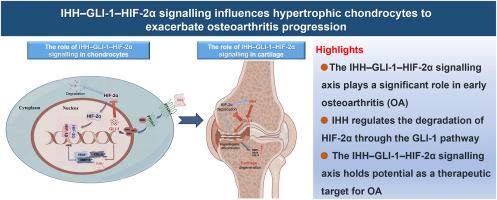
Abstract Image

IHH-GLI-1-HIF-2α信号影响肥大软骨细胞,加剧骨关节炎进展
背景软骨细胞肥大是骨关节炎(OA)治疗的一个潜在靶点,印度刺猬(IHH)、胶质瘤相关癌基因同源物(GLI)和缺氧诱导因子-2α(HIF-2α)与OA进展过程中的软骨细胞肥大密切相关。虽然 IHH 可以调节软骨细胞肥大,但干扰 IHH 信号并不能达到预期的治疗效果,而且还存在安全隐患,因此有必要进一步阐明 IHH 影响关节软骨退化的具体机制。抑制软骨中HIF-2α的过度表达可减缓早期OA的进展,但HIF-2α在OA软骨中积累的机制仍不清楚。本研究的目的是基于早期骨关节炎(OA)小鼠模型和体外软骨细胞模型,确定 Ihh 及其下游因子在软骨细胞中的功能。方法研究 IHH-GLI-1 通路在正常和早期退化人类软骨中的表达水平和位置,并与 HIF-2α 及其下游因子进行比较。通过 RT-qPCR、Western 印迹、水晶紫染色和 EdU 检测,评估了 IHH-GLI-1-HIF-2α 信号轴在正常软骨细胞和炎症条件下软骨细胞中的特殊调控机制。验证了 IHH 对早期软骨退化的影响,以及 IHH-GLI-1 通路与 Col2a1-CreERT2;Ihhfl/fl 小鼠中 HIF-2α 及其下游因子的表达水平和表达位置之间的关系。在体外,生理缺氧条件下软骨细胞中的 IHH-GLI-1-HIF-2α 信号激活会抑制软骨细胞的增殖。在受到炎症环境刺激的软骨细胞中,IHH 通过 GLI-1 通路抑制了 HIF-2α 的降解,从而促进了 HIF-2α 蛋白的表达。HIF-2α表达的升高进一步提高了细胞内IHH-GLI-1的水平,形成了一个正反馈回路,共同调节下游肥大因子和基质降解因子的表达。在体内,小鼠软骨细胞中条件性 Ihh 基因敲除可下调早期退行性软骨组织中 Hif-2α 蛋白的表达,并影响下游 Vegf 和肥大因子的表达。结论在OA进展过程中,IHH-GLI-1-HIF-2α轴主要在肥大软骨细胞内发挥作用,通过调控肥大软骨细胞功能、软骨基质降解和微血管侵袭加剧软骨退行性变。
本文章由计算机程序翻译,如有差异,请以英文原文为准。
求助全文
约1分钟内获得全文 求助全文
来源期刊
Journal of Orthopaedic Translation
Journal of Orthopaedic Translation Medicine-Orthopedics and Sports Medicine
CiteScore
11.80
自引率
13.60%
发文量
91
审稿时长
29 days
期刊介绍: The Journal of Orthopaedic Translation (JOT) is the official peer-reviewed, open access journal of the Chinese Speaking Orthopaedic Society (CSOS) and the International Chinese Musculoskeletal Research Society (ICMRS). It is published quarterly, in January, April, July and October, by Elsevier.
×
引用
GB/T 7714-2015
复制
MLA
复制
APA
复制
导出至
BibTeX EndNote RefMan NoteFirst NoteExpress
×
提示
您的信息不完整,为了账户安全,请先补充。
现在去补充
×
提示
您因"违规操作"
具体请查看互助需知
我知道了
×
提示
确定
请完成安全验证×
copy
已复制链接
快去分享给好友吧!
我知道了
右上角分享
点击右上角分享
0
联系我们:info@booksci.cn Book学术提供免费学术资源搜索服务,方便国内外学者检索中英文文献。致力于提供最便捷和优质的服务体验。 Copyright © 2023 布克学术 All rights reserved.
京ICP备2023020795号-1
ghs 京公网安备 11010802042870号
Book学术文献互助
Book学术文献互助群
群 号:481959085
Book学术官方微信