BreasTDLUSeg: A coarse-to-fine framework for segmentation of breast terminal duct lobular units on histopathological whole-slide images
IF 5.4
2区 医学
Q1 ENGINEERING, BIOMEDICAL
Computerized Medical Imaging and Graphics
Pub Date : 2024-09-19
DOI:10.1016/j.compmedimag.2024.102432
引用次数: 0
Abstract
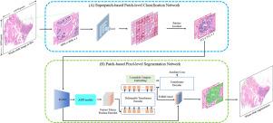
Automatic segmentation of breast terminal duct lobular units (TDLUs) on histopathological whole-slide images (WSIs) is crucial for the quantitative evaluation of TDLUs in the diagnostic and prognostic analysis of breast cancer. However, TDLU segmentation remains a great challenge due to its highly heterogeneous sizes, structures, and morphologies as well as the small areas on WSIs. In this study, we propose BreasTDLUSeg, an efficient coarse-to-fine two-stage framework based on multi-scale attention to achieve localization and precise segmentation of TDLUs on hematoxylin and eosin (H&E)-stained WSIs. BreasTDLUSeg consists of two networks: a superpatch-based patch-level classification network (SPPC-Net) and a patch-based pixel-level segmentation network (PPS-Net). SPPC-Net takes a superpatch as input and adopts a sub-region classification head to classify each patch within the superpatch as TDLU positive or negative. PPS-Net takes the TDLU positive patches derived from SPPC-Net as input. PPS-Net deploys a multi-scale CNN-Transformer as an encoder to learn enhanced multi-scale morphological representations and an upsampler to generate pixel-wise segmentation masks for the TDLU positive patches. We also constructed two breast cancer TDLU datasets containing a total of 530 superpatch images with patch-level annotations and 2322 patch images with pixel-level annotations to enable the development of TDLU segmentation methods. Experiments on the two datasets demonstrate that BreasTDLUSeg outperforms other state-of-the-art methods with the highest Dice similarity coefficients of 79.97% and 92.93%, respectively. The proposed method shows great potential to assist pathologists in the pathological analysis of breast cancer. An open-source implementation of our approach can be found at https://github.com/Dian-kai/BreasTDLUSeg.

BreasTDLUSeg:在组织病理学全切片图像上分割乳腺末端导管小叶单元的粗到细框架。
在组织病理全切片图像(WSI)上自动分割乳腺末端导管小叶单位(TDLU)对于乳腺癌诊断和预后分析中定量评估 TDLU 至关重要。然而,由于 TDLU 的大小、结构和形态差异很大,而且在 WSIs 上的面积很小,因此 TDLU 的分割仍然是一项巨大的挑战。在本研究中,我们提出了基于多尺度关注的高效粗到细两阶段框架 BreasTDLUSeg,以实现苏木精和伊红(H&E)染色 WSI 上 TDLU 的定位和精确分割。BreasTDLUSeg 由两个网络组成:基于超斑的斑块级分类网络(SPPC-Net)和基于斑块的像素级分割网络(PPS-Net)。SPPC-Net 以超级斑块为输入,采用子区域分类头将超级斑块中的每个斑块划分为 TDLU 正片或负片。PPS-Net 将 SPPC-Net 得出的 TDLU 正片作为输入。PPS-Net 部署了一个多尺度 CNN 变换器作为编码器,以学习增强的多尺度形态学表示,并部署了一个上采样器,为 TDLU 阳性斑块生成像素分割掩码。我们还构建了两个乳腺癌 TDLU 数据集,共包含 530 幅带有斑块级注释的超级斑块图像和 2322 幅带有像素级注释的斑块图像,以便开发 TDLU 分割方法。在这两个数据集上的实验表明,BreasTDLUSeg 优于其他最先进的方法,其最高 Dice 相似系数分别为 79.97% 和 92.93%。所提出的方法在协助病理学家进行乳腺癌病理分析方面显示出巨大的潜力。我们的方法的开源实现可以在 https://github.com/Dian-kai/BreasTDLUSeg 上找到。
本文章由计算机程序翻译,如有差异,请以英文原文为准。
求助全文
约1分钟内获得全文
求助全文
来源期刊
CiteScore
10.70
自引率
3.50%
发文量
71
审稿时长
26 days
期刊介绍:
The purpose of the journal Computerized Medical Imaging and Graphics is to act as a source for the exchange of research results concerning algorithmic advances, development, and application of digital imaging in disease detection, diagnosis, intervention, prevention, precision medicine, and population health. Included in the journal will be articles on novel computerized imaging or visualization techniques, including artificial intelligence and machine learning, augmented reality for surgical planning and guidance, big biomedical data visualization, computer-aided diagnosis, computerized-robotic surgery, image-guided therapy, imaging scanning and reconstruction, mobile and tele-imaging, radiomics, and imaging integration and modeling with other information relevant to digital health. The types of biomedical imaging include: magnetic resonance, computed tomography, ultrasound, nuclear medicine, X-ray, microwave, optical and multi-photon microscopy, video and sensory imaging, and the convergence of biomedical images with other non-imaging datasets.

 求助内容:
求助内容: 应助结果提醒方式:


