Hakan Özdemir, Furkan Kırık, Gizem Elif Atlı, Begüm Petek Al
{"title":"Multilayered Inverted Internal Limiting Membrane Flap Technique in Optic Disc Pit Maculopathy.","authors":"Hakan Özdemir, Furkan Kırık, Gizem Elif Atlı, Begüm Petek Al","doi":"10.4274/tjo.galenos.2024.46402","DOIUrl":null,"url":null,"abstract":"<p><strong>Objectives: </strong>To evaluate the anatomical and visual outcomes of the multilayered inverted internal limiting membrane (ML-ILM) flap technique in the treatment of optic disc pit maculopathy (ODPM).</p><p><strong>Materials and methods: </strong>In this retrospective interventional case series, medical records and macular spectral-domain optical coherence tomography images of patients who underwent combined pars plana vitrectomy with ML-ILM flap surgery for ODPM were analyzed. Best-corrected visual acuity (BCVA) and central macular thickness (CMT) at postoperative 6 months were compared with baseline findings. Intraoperative and postoperative complications, fluid resolution time, and recurrence during follow-up were recorded.</p><p><strong>Results: </strong>Five eyes of 5 patients with ODPM were included in the study. According to the preoperative macular fluid characteristics, 2 patients had only intraretinal fluid, while 3 patients had intraretinal and subretinal fluid. The preoperative median BCVA was 1.0 logarithm of the minimum angle of resolution (logMAR) (range, 1.0-1.3 logMAR), and the CMT was 560 μm (range, 452-667 μm). At the 6-month postoperative follow-up, the median BCVA was 0.40 logMAR (range, 0.1-0.7 logMAR), and CMT was 315 μm (range, 265-326 μm) (p=0.042 and p=0.043, respectively). During the 6-month follow-up period, no recurrence or full-thickness macular hole formation was observed.</p><p><strong>Conclusion: </strong>The ML-ILM flap technique is a preferable surgical option to achieve both high anatomical and functional success and flap stabilization.</p>","PeriodicalId":23373,"journal":{"name":"Turkish Journal of Ophthalmology","volume":"54 5","pages":"275-281"},"PeriodicalIF":0.0000,"publicationDate":"2024-10-25","publicationTypes":"Journal Article","fieldsOfStudy":null,"isOpenAccess":false,"openAccessPdf":"https://www.ncbi.nlm.nih.gov/pmc/articles/PMC11589234/pdf/","citationCount":"0","resultStr":null,"platform":"Semanticscholar","paperid":null,"PeriodicalName":"Turkish Journal of Ophthalmology","FirstCategoryId":"1085","ListUrlMain":"https://doi.org/10.4274/tjo.galenos.2024.46402","RegionNum":0,"RegionCategory":null,"ArticlePicture":[],"TitleCN":null,"AbstractTextCN":null,"PMCID":null,"EPubDate":"","PubModel":"","JCR":"Q3","JCRName":"Medicine","Score":null,"Total":0}
引用次数: 0
Abstract
Objectives: To evaluate the anatomical and visual outcomes of the multilayered inverted internal limiting membrane (ML-ILM) flap technique in the treatment of optic disc pit maculopathy (ODPM).
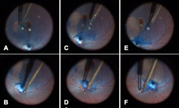
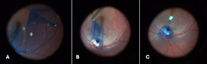
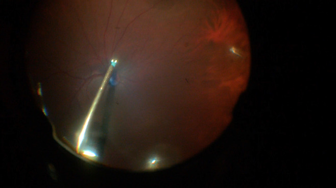
Materials and methods: In this retrospective interventional case series, medical records and macular spectral-domain optical coherence tomography images of patients who underwent combined pars plana vitrectomy with ML-ILM flap surgery for ODPM were analyzed. Best-corrected visual acuity (BCVA) and central macular thickness (CMT) at postoperative 6 months were compared with baseline findings. Intraoperative and postoperative complications, fluid resolution time, and recurrence during follow-up were recorded.
Results: Five eyes of 5 patients with ODPM were included in the study. According to the preoperative macular fluid characteristics, 2 patients had only intraretinal fluid, while 3 patients had intraretinal and subretinal fluid. The preoperative median BCVA was 1.0 logarithm of the minimum angle of resolution (logMAR) (range, 1.0-1.3 logMAR), and the CMT was 560 μm (range, 452-667 μm). At the 6-month postoperative follow-up, the median BCVA was 0.40 logMAR (range, 0.1-0.7 logMAR), and CMT was 315 μm (range, 265-326 μm) (p=0.042 and p=0.043, respectively). During the 6-month follow-up period, no recurrence or full-thickness macular hole formation was observed.
Conclusion: The ML-ILM flap technique is a preferable surgical option to achieve both high anatomical and functional success and flap stabilization.
期刊介绍:
The Turkish Journal of Ophthalmology (TJO) is the only scientific periodical publication of the Turkish Ophthalmological Association and has been published since January 1929. In its early years, the journal was published in Turkish and French. Although there were temporary interruptions in the publication of the journal due to various challenges, the Turkish Journal of Ophthalmology has been published continually from 1971 to the present. The target audience includes specialists and physicians in training in ophthalmology in all relevant disciplines.

 求助内容:
求助内容: 应助结果提醒方式:


