Correlative Raman spectroscopy and electron microscopy identifies glycogen rich deposits correlated with local structural defects in long bones of type IV osteogenesis imperfecta patients
IF 2.7
3区 生物学
Q3 BIOCHEMISTRY & MOLECULAR BIOLOGY
引用次数: 0
Abstract
Osteogenesis imperfecta (OI) is a genetic bone disease occurring in approximately 1 in 10,000 births, usually as a result of genetic mutation. OI patients suffer from increased fracture risk and – depending on the severity of the disease – deformation of the limbs, which can even lead to perinatal death.
Despite extensive studies, the way in which the genetic mutation is translated into structural and compositional anomalies of the tissue is still an open question. Different observations have been reported, ranging from no structural (or chemical) differences to completely chaotic bone structure and composition.
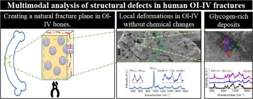
Here, we investigated bone samples from two adolescent OI-IV patients, focusing on the bone structure and chemistry in naturally occurring fractures. The exposed fracture plane allows the investigation of the structure and composition of the weakest bone plane. We do so by combining scanning electron microscopy (SEM) imaging with chemical information from Raman microscopy.
The exposed fracture planes show different regions within the same tissue, displaying normal osteonal structures next to disorganized osteons and totally disordered structures, while the collagen mineralization in all cases is similar to that of a healthy bone.
In addition, we also detected significant amounts of depositions of glycogen-rich, organic, globules of 250–1000 nm in size. These depositions point to a role of cellular disfunction in the disorganization of the collagen in qualitative OI.
Overall, our results unite multiple, sometimes contradicting views from the literature on qualitative OI.

拉曼光谱和电子显微镜的相关研究发现,富含糖原的沉积物与 IV 型成骨不全症患者长骨的局部结构缺陷有关。
成骨不全症(OI)是一种遗传性骨病,大约每 1 万名新生儿中就有 1 人患有这种疾病,通常是由于基因突变造成的。成骨不全症患者骨折风险增加,根据病情严重程度,四肢会变形,甚至导致围产期死亡。尽管进行了大量研究,但基因突变如何转化为组织结构和组成异常仍是一个未决问题。从无结构(或化学)差异到完全混乱的骨结构和组成,不同的观察结果均有报道。在此,我们研究了两名青少年 OI-IV 患者的骨样本,重点是自然发生骨折时的骨结构和化学成分。通过暴露的骨折面可以研究最薄弱骨面的结构和成分。为此,我们将扫描电子显微镜(SEM)成像与拉曼显微镜的化学信息相结合。裸露的断裂面显示了同一组织内的不同区域,有正常的骨细胞结构,也有杂乱无章的骨细胞和完全无序的结构,而所有情况下的胶原矿化都与健康骨骼相似。此外,我们还检测到大量富含糖原的有机球状沉积物,大小为 250-1000 纳米。这些沉积物表明,在定性 OI 中,细胞功能失调在胶原组织混乱中起了作用。总之,我们的研究结果将定性 OI 文献中多种有时相互矛盾的观点统一起来。
本文章由计算机程序翻译,如有差异,请以英文原文为准。
求助全文
约1分钟内获得全文
求助全文
来源期刊

Journal of structural biology
生物-生化与分子生物学
CiteScore
6.30
自引率
3.30%
发文量
88
审稿时长
65 days
期刊介绍:
Journal of Structural Biology (JSB) has an open access mirror journal, the Journal of Structural Biology: X (JSBX), sharing the same aims and scope, editorial team, submission system and rigorous peer review. Since both journals share the same editorial system, you may submit your manuscript via either journal homepage. You will be prompted during submission (and revision) to choose in which to publish your article. The editors and reviewers are not aware of the choice you made until the article has been published online. JSB and JSBX publish papers dealing with the structural analysis of living material at every level of organization by all methods that lead to an understanding of biological function in terms of molecular and supermolecular structure.
Techniques covered include:
• Light microscopy including confocal microscopy
• All types of electron microscopy
• X-ray diffraction
• Nuclear magnetic resonance
• Scanning force microscopy, scanning probe microscopy, and tunneling microscopy
• Digital image processing
• Computational insights into structure
 求助内容:
求助内容: 应助结果提醒方式:


