Extracapsular extension risk assessment using an artificial intelligence prostate cancer mapping algorithm

IF 1.6 Q3 UROLOGY & NEPHROLOGY
BJUI compass Pub Date : 2024-08-26 DOI:10.1002/bco2.421
Alan Priester, Sakina Mohammed Mota, Kyla P. Grunden, Joshua Shubert, Shannon Richardson, Anthony Sisk, Ely R. Felker, James Sayre, Leonard S. Marks, Shyam Natarajan, Wayne G. Brisbane
{"title":"Extracapsular extension risk assessment using an artificial intelligence prostate cancer mapping algorithm","authors":"Alan Priester,&nbsp;Sakina Mohammed Mota,&nbsp;Kyla P. Grunden,&nbsp;Joshua Shubert,&nbsp;Shannon Richardson,&nbsp;Anthony Sisk,&nbsp;Ely R. Felker,&nbsp;James Sayre,&nbsp;Leonard S. Marks,&nbsp;Shyam Natarajan,&nbsp;Wayne G. Brisbane","doi":"10.1002/bco2.421","DOIUrl":null,"url":null,"abstract":"<div>\n \n \n <section>\n \n <h3> Objective</h3>\n \n <p>The objective of this study is to compare detection rates of extracapsular extension (ECE) of prostate cancer (PCa) using artificial intelligence (AI)-generated cancer maps versus MRI and conventional nomograms.</p>\n </section>\n \n <section>\n \n <h3> Materials and methods</h3>\n \n <p>We retrospectively analysed data from 147 patients who received MRI-targeted biopsy and subsequent radical prostatectomy between September 2016 and May 2022. AI-based software cleared by the United States Food and Drug Administration (Unfold AI, Avenda Health) was used to map 3D cancer probability and estimate ECE risk. Conventional ECE predictors including MRI Likert scores, capsular contact length of MRI-visible lesions, PSMA T stage, Partin tables, and the “PRedicting ExtraCapsular Extension” nomogram were used for comparison.</p>\n \n <p>Postsurgical specimens were processed using whole-mount histopathology sectioning, and a genitourinary pathologist assessed each quadrant for ECE presence. ECE predictors were then evaluated on the patient (Unfold AI versus all comparators) and quadrant level (Unfold AI versus MRI Likert score). Receiver operator characteristic curves were generated and compared using DeLong's test.</p>\n </section>\n \n <section>\n \n <h3> Results</h3>\n \n <p>Unfold AI had a significantly higher area under the curve (AUC = 0.81) than other predictors for patient-level ECE prediction. Unfold AI achieved 68% sensitivity, 78% specificity, 71% positive predictive value, and 75% negative predictive value. At the quadrant level, Unfold AI exceeded the AUC of MRI Likert scores for posterior (0.89 versus 0.82, <i>p</i> = 0.003), anterior (0.84 versus 0.80, <i>p</i> = 0.34), and all quadrants (0.89 versus 0.82, <i>p</i> = 0.002). The false negative rate of Unfold AI was lower than MRI in both the anterior (−60%) and posterior prostate (−40%).</p>\n </section>\n \n <section>\n \n <h3> Conclusions</h3>\n \n <p>Unfold AI accurately predicted ECE risk, outperforming conventional methodologies. It notably improved ECE prediction over MRI in posterior quadrants, with the potential to inform nerve-spare technique and prevent positive margins. By enhancing PCa staging and risk stratification, AI-based cancer mapping may lead to better oncological and functional outcomes for patients.</p>\n </section>\n </div>","PeriodicalId":72420,"journal":{"name":"BJUI compass","volume":"5 10","pages":"986-997"},"PeriodicalIF":1.6000,"publicationDate":"2024-08-26","publicationTypes":"Journal Article","fieldsOfStudy":null,"isOpenAccess":false,"openAccessPdf":"https://onlinelibrary.wiley.com/doi/epdf/10.1002/bco2.421","citationCount":"0","resultStr":null,"platform":"Semanticscholar","paperid":null,"PeriodicalName":"BJUI compass","FirstCategoryId":"1085","ListUrlMain":"https://onlinelibrary.wiley.com/doi/10.1002/bco2.421","RegionNum":0,"RegionCategory":null,"ArticlePicture":[],"TitleCN":null,"AbstractTextCN":null,"PMCID":null,"EPubDate":"","PubModel":"","JCR":"Q3","JCRName":"UROLOGY & NEPHROLOGY","Score":null,"Total":0}
引用次数: 0

Abstract

Objective

The objective of this study is to compare detection rates of extracapsular extension (ECE) of prostate cancer (PCa) using artificial intelligence (AI)-generated cancer maps versus MRI and conventional nomograms.

Materials and methods

We retrospectively analysed data from 147 patients who received MRI-targeted biopsy and subsequent radical prostatectomy between September 2016 and May 2022. AI-based software cleared by the United States Food and Drug Administration (Unfold AI, Avenda Health) was used to map 3D cancer probability and estimate ECE risk. Conventional ECE predictors including MRI Likert scores, capsular contact length of MRI-visible lesions, PSMA T stage, Partin tables, and the “PRedicting ExtraCapsular Extension” nomogram were used for comparison.

Postsurgical specimens were processed using whole-mount histopathology sectioning, and a genitourinary pathologist assessed each quadrant for ECE presence. ECE predictors were then evaluated on the patient (Unfold AI versus all comparators) and quadrant level (Unfold AI versus MRI Likert score). Receiver operator characteristic curves were generated and compared using DeLong's test.

Results

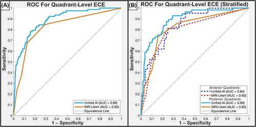
Unfold AI had a significantly higher area under the curve (AUC = 0.81) than other predictors for patient-level ECE prediction. Unfold AI achieved 68% sensitivity, 78% specificity, 71% positive predictive value, and 75% negative predictive value. At the quadrant level, Unfold AI exceeded the AUC of MRI Likert scores for posterior (0.89 versus 0.82, p = 0.003), anterior (0.84 versus 0.80, p = 0.34), and all quadrants (0.89 versus 0.82, p = 0.002). The false negative rate of Unfold AI was lower than MRI in both the anterior (−60%) and posterior prostate (−40%).

Conclusions

Unfold AI accurately predicted ECE risk, outperforming conventional methodologies. It notably improved ECE prediction over MRI in posterior quadrants, with the potential to inform nerve-spare technique and prevent positive margins. By enhancing PCa staging and risk stratification, AI-based cancer mapping may lead to better oncological and functional outcomes for patients.
Abstract Image

利用人工智能前列腺癌映射算法进行囊外扩展风险评估
目的 本研究旨在比较使用人工智能(AI)生成的癌症图谱与核磁共振成像和传统提名图对前列腺癌(PCa)囊外扩展(ECE)的检测率。 材料与方法 我们回顾性分析了2016年9月至2022年5月期间接受磁共振成像靶向活检并随后接受根治性前列腺切除术的147名患者的数据。我们使用经美国食品药品管理局批准的人工智能软件(Unfold AI,Avenda Health)绘制三维癌症概率图并估算 ECE 风险。传统的ECE预测指标包括MRI Likert评分、MRI可见病灶的囊接触长度、PSMA T分期、Partin表和 "预测囊外扩展 "提名图,用于比较。 手术后标本采用全贴面组织病理学切片法进行处理,由泌尿生殖系统病理学家评估每个象限是否存在 ECE。然后对患者(Unfold AI 与所有比较者)和象限水平(Unfold AI 与 MRI Likert 评分)的 ECE 预测因子进行评估。生成接收者操作者特征曲线,并使用 DeLong 检验进行比较。 结果 在预测患者层面的 ECE 时,Unfold AI 的曲线下面积(AUC = 0.81)明显高于其他预测指标。Unfold AI 的灵敏度为 68%,特异性为 78%,阳性预测值为 71%,阴性预测值为 75%。在象限水平上,Unfold AI 在后部(0.89 对 0.82,p = 0.003)、前部(0.84 对 0.80,p = 0.34)和所有象限(0.89 对 0.82,p = 0.002)的 AUC 超过了 MRI Likert 评分。在前列腺前部(-60%)和后部(-40%),Unfold AI 的假阴性率均低于 MRI。 结论 Unfold AI 能准确预测 ECE 风险,优于传统方法。与核磁共振成像相比,它显著提高了后象限的ECE预测能力,有可能为神经剥离技术提供参考,并防止出现阳性边缘。通过加强 PCa 分期和风险分层,基于人工智能的癌症图谱可能会为患者带来更好的肿瘤和功能治疗效果。
本文章由计算机程序翻译,如有差异,请以英文原文为准。
求助全文
约1分钟内获得全文 求助全文
来源期刊
CiteScore
2.30
自引率
0.00%
发文量
0
审稿时长
12 weeks
×
引用
GB/T 7714-2015
复制
MLA
复制
APA
复制
导出至
BibTeX EndNote RefMan NoteFirst NoteExpress
×
提示
您的信息不完整,为了账户安全,请先补充。
现在去补充
×
提示
您因"违规操作"
具体请查看互助需知
我知道了
×
提示
确定
请完成安全验证×
copy
已复制链接
快去分享给好友吧!
我知道了
右上角分享
点击右上角分享
0
联系我们:info@booksci.cn Book学术提供免费学术资源搜索服务,方便国内外学者检索中英文文献。致力于提供最便捷和优质的服务体验。 Copyright © 2023 布克学术 All rights reserved.
京ICP备2023020795号-1
ghs 京公网安备 11010802042870号
Book学术文献互助
Book学术文献互助群
群 号:481959085
Book学术官方微信