{"title":"Medial soft tissue release is also related to the anterior stability of cruciate-retaining total knee arthroplasty: a cadaveric study.","authors":"Sayako Sakai, Shinichiro Nakamura, Takahiro Maeda, Shinichi Kuriyama, Kohei Nishitani, Yugo Morita, Yugo Morita, Yusuke Yamawaki, Yuki Shinya, Shuichi Matsuda","doi":"10.1186/s43019-024-00233-6","DOIUrl":null,"url":null,"abstract":"<p><strong>Background: </strong>Medial soft tissue release is occasionally performed to achieve mediolateral ligament balance in total knee arthroplasty (TKA), whose sequential effect on mediolateral and anteroposterior stability remains unclear. This study aimed to quantitatively evaluate the difference in mediolateral and anteroposterior stability according to a sequential medial soft tissue release in TKA.</p><p><strong>Methods: </strong>Cruciate-retaining TKA was performed in six cadaveric knees. Medial and lateral joint gaps, varus-valgus angle, and tibial anterior and posterior translations relative to the femur with pulling and pushing forces, respectively, were measured. All measurements were performed at full extension and 45° and 90° flexion after release of the deep medial collateral ligament (MCL) (stage 1), the posteromedial capsule (stage 2), and the superficial MCL (stage 3). Mediolateral and anteroposterior stability were compared between stages, and correlations between mediolateral and anteroposterior stability were analyzed.</p><p><strong>Results: </strong>Medial joint gap significantly increased from stages 1 to 3 by 3.2 mm, 6.8 mm, and 7.2 mm at extension, 45° flexion, and 90° flexion, respectively, and from stages 2 to 3 by 3.5 mm at extension. Varus-valgus angle was varus at stage 2, which turned to valgus at stage 3 (-2.7° to 0.8°, -2.2° to 4.3°, and -5.5° to 2.5° at extension, 45° flexion, and 90° flexion, respectively). Anterior translation at 90° flexion significantly increased from stages 1 and 2 to stage 3 by 11.5 mm and 8.2 mm, respectively, which was significantly correlated with medial gap (r = 0.681) and varus-valgus angle (r = 0.495).</p><p><strong>Conclusions: </strong>Medial soft tissue release also increased tibial anterior translation as well as medial joint gap, and medial joint gap and tibial anterior translation were significantly correlated. Surgeons should be careful not to create too large medial joint gap and tibial anterior translation in flexion by excessive medial release up to the superficial MCL for achieving an equal mediolateral joint gap in extension.</p>","PeriodicalId":36317,"journal":{"name":"Knee Surgery and Related Research","volume":"36 1","pages":"29"},"PeriodicalIF":4.4000,"publicationDate":"2024-10-08","publicationTypes":"Journal Article","fieldsOfStudy":null,"isOpenAccess":false,"openAccessPdf":"https://www.ncbi.nlm.nih.gov/pmc/articles/PMC11459880/pdf/","citationCount":"0","resultStr":null,"platform":"Semanticscholar","paperid":null,"PeriodicalName":"Knee Surgery and Related Research","FirstCategoryId":"1085","ListUrlMain":"https://doi.org/10.1186/s43019-024-00233-6","RegionNum":0,"RegionCategory":null,"ArticlePicture":[],"TitleCN":null,"AbstractTextCN":null,"PMCID":null,"EPubDate":"","PubModel":"","JCR":"Q2","JCRName":"Medicine","Score":null,"Total":0}
引用次数: 0
Abstract
Background: Medial soft tissue release is occasionally performed to achieve mediolateral ligament balance in total knee arthroplasty (TKA), whose sequential effect on mediolateral and anteroposterior stability remains unclear. This study aimed to quantitatively evaluate the difference in mediolateral and anteroposterior stability according to a sequential medial soft tissue release in TKA.
Methods: Cruciate-retaining TKA was performed in six cadaveric knees. Medial and lateral joint gaps, varus-valgus angle, and tibial anterior and posterior translations relative to the femur with pulling and pushing forces, respectively, were measured. All measurements were performed at full extension and 45° and 90° flexion after release of the deep medial collateral ligament (MCL) (stage 1), the posteromedial capsule (stage 2), and the superficial MCL (stage 3). Mediolateral and anteroposterior stability were compared between stages, and correlations between mediolateral and anteroposterior stability were analyzed.
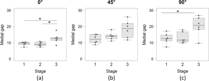
Results: Medial joint gap significantly increased from stages 1 to 3 by 3.2 mm, 6.8 mm, and 7.2 mm at extension, 45° flexion, and 90° flexion, respectively, and from stages 2 to 3 by 3.5 mm at extension. Varus-valgus angle was varus at stage 2, which turned to valgus at stage 3 (-2.7° to 0.8°, -2.2° to 4.3°, and -5.5° to 2.5° at extension, 45° flexion, and 90° flexion, respectively). Anterior translation at 90° flexion significantly increased from stages 1 and 2 to stage 3 by 11.5 mm and 8.2 mm, respectively, which was significantly correlated with medial gap (r = 0.681) and varus-valgus angle (r = 0.495).
Conclusions: Medial soft tissue release also increased tibial anterior translation as well as medial joint gap, and medial joint gap and tibial anterior translation were significantly correlated. Surgeons should be careful not to create too large medial joint gap and tibial anterior translation in flexion by excessive medial release up to the superficial MCL for achieving an equal mediolateral joint gap in extension.

 求助内容:
求助内容: 应助结果提醒方式:


