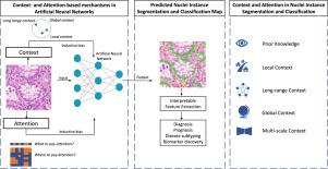
A survey on cell nuclei instance segmentation and classification: Leveraging context and attention
IF 10.7
1区 医学
Q1 COMPUTER SCIENCE, ARTIFICIAL INTELLIGENCE
引用次数: 0
Abstract
Nuclear-derived morphological features and biomarkers provide relevant insights regarding the tumour microenvironment, while also allowing diagnosis and prognosis in specific cancer types. However, manually annotating nuclei from the gigapixel Haematoxylin and Eosin (H&E)-stained Whole Slide Images (WSIs) is a laborious and costly task, meaning automated algorithms for cell nuclei instance segmentation and classification could alleviate the workload of pathologists and clinical researchers and at the same time facilitate the automatic extraction of clinically interpretable features for artificial intelligence (AI) tools. But due to high intra- and inter-class variability of nuclei morphological and chromatic features, as well as H&E-stains susceptibility to artefacts, state-of-the-art algorithms cannot correctly detect and classify instances with the necessary performance. In this work, we hypothesize context and attention inductive biases in artificial neural networks (ANNs) could increase the performance and generalization of algorithms for cell nuclei instance segmentation and classification. To understand the advantages, use-cases, and limitations of context and attention-based mechanisms in instance segmentation and classification, we start by reviewing works in computer vision and medical imaging. We then conduct a thorough survey on context and attention methods for cell nuclei instance segmentation and classification from H&E-stained microscopy imaging, while providing a comprehensive discussion of the challenges being tackled with context and attention. Besides, we illustrate some limitations of current approaches and present ideas for future research. As a case study, we extend both a general (Mask-RCNN) and a customized (HoVer-Net) instance segmentation and classification methods with context- and attention-based mechanisms and perform a comparative analysis on a multicentre dataset for colon nuclei identification and counting.
Although pathologists rely on context at multiple levels while paying attention to specific Regions of Interest (RoIs) when analysing and annotating WSIs, our findings suggest translating that domain knowledge into algorithm design is no trivial task, but to fully exploit these mechanisms in ANNs, the scientific understanding of these methods should first be addressed.

细胞核实例分割和分类调查:利用上下文和注意力
核源性形态特征和生物标记物能提供有关肿瘤微环境的相关信息,同时还能对特定癌症类型进行诊断和预后判断。然而,从千百万像素的血苏木精和伊红(H&E)染色的全切片图像(WSI)中手动标注细胞核是一项费力又费钱的工作,这意味着细胞核实例分割和分类的自动化算法可以减轻病理学家和临床研究人员的工作量,同时有助于为人工智能(AI)工具自动提取临床上可解释的特征。但是,由于细胞核形态和色度特征在类内和类间的高变异性,以及 H&E 染色易受伪影影响,最先进的算法无法以必要的性能正确检测和分类实例。在这项工作中,我们假设人工神经网络(ANN)中的上下文和注意力归纳偏差可以提高细胞核实例分割和分类算法的性能和泛化。为了了解基于上下文和注意力的机制在实例分割和分类中的优势、用例和局限性,我们首先回顾了计算机视觉和医学成像领域的研究成果。然后,我们深入研究了从 H&E 染色显微镜成像中进行细胞核实例分割和分类的上下文和注意力方法,同时对上下文和注意力所面临的挑战进行了全面讨论。此外,我们还说明了当前方法的一些局限性,并提出了未来研究的思路。作为一项案例研究,我们利用基于上下文和注意力的机制扩展了通用(Mask-RCNN)和定制(HoVer-Net)实例分割和分类方法,并在多中心数据集上对结肠核的识别和计数进行了比较分析。虽然病理学家在分析和注释 WSIs 时依赖于多层次的上下文,同时关注特定的感兴趣区(RoIs),但我们的研究结果表明,将领域知识转化为算法设计并非易事,但要在人工神经网络中充分利用这些机制,首先应解决对这些方法的科学理解问题。
本文章由计算机程序翻译,如有差异,请以英文原文为准。
求助全文
约1分钟内获得全文
求助全文
来源期刊

Medical image analysis
工程技术-工程:生物医学
CiteScore
22.10
自引率
6.40%
发文量
309
审稿时长
6.6 months
期刊介绍:
Medical Image Analysis serves as a platform for sharing new research findings in the realm of medical and biological image analysis, with a focus on applications of computer vision, virtual reality, and robotics to biomedical imaging challenges. The journal prioritizes the publication of high-quality, original papers contributing to the fundamental science of processing, analyzing, and utilizing medical and biological images. It welcomes approaches utilizing biomedical image datasets across all spatial scales, from molecular/cellular imaging to tissue/organ imaging.
 求助内容:
求助内容: 应助结果提醒方式:


