Structural analysis of microtubule binding by minus-end targeting protein Spiral2

IF 4.6 2区 生物学 Q1 BIOCHEMISTRY & MOLECULAR BIOLOGY
Marina Ohno , Yuuki Higuchi , Kazune Yamai , Sotaro Fuchigami , Takema Sasaki , Yoshihisa Oda , Ikuko Hayashi
{"title":"Structural analysis of microtubule binding by minus-end targeting protein Spiral2","authors":"Marina Ohno ,&nbsp;Yuuki Higuchi ,&nbsp;Kazune Yamai ,&nbsp;Sotaro Fuchigami ,&nbsp;Takema Sasaki ,&nbsp;Yoshihisa Oda ,&nbsp;Ikuko Hayashi","doi":"10.1016/j.bbamcr.2024.119858","DOIUrl":null,"url":null,"abstract":"<div><div>Microtubules (MTs) are dynamic cytoskeletal polymers that play a critical role in determining cell polarity and shape. In plant cells, acentrosomal MTs are localized on the cell surface and are referred to as cortical MTs. Cortical MTs nucleate in the cell cortex and detach from nucleation sites. The released MT filaments perform treadmilling, with the plus-ends of MTs polymerizing and the minus-ends depolymerizing. Minus-end targeting proteins, -TIPs, include Spiral2, which regulates the minus-end dynamics of acentrosomal MTs. Spiral2 accumulates autonomously at MT minus-ends and inhibits filament shrinkage, but the mechanism by which Spiral2 specifically recognizes minus-ends of MTs remains unknown. Here we describe the crystal structure of Spiral2's N-terminal MT-binding domain. The structural properties of this domain resemble those of the HEAT repeat structure of the tumor overexpressed gene (TOG) domain, but the number of HEAT repeats is different and the conformation is highly arched. Gel filtration and co-sedimentation analyses demonstrate that the domain binds preferentially to MT filaments rather than the tubulin dimer, and that the tubulin-binding mode of Spiral2 <em>via</em> the basic surface is similar to that of the TOG domain. We constructed an <em>in silico</em> model of the Spiral2-tubulin complex to identify residues that potentially recognize tubulin. Mutational analysis revealed that the key residues inferred in the model are involved in microtubule recognition, and provide insight into the mechanism by which end-targeting proteins stabilize MT ends.</div></div>","PeriodicalId":8754,"journal":{"name":"Biochimica et biophysica acta. Molecular cell research","volume":"1871 8","pages":"Article 119858"},"PeriodicalIF":4.6000,"publicationDate":"2024-10-04","publicationTypes":"Journal Article","fieldsOfStudy":null,"isOpenAccess":false,"openAccessPdf":"","citationCount":"0","resultStr":null,"platform":"Semanticscholar","paperid":null,"PeriodicalName":"Biochimica et biophysica acta. Molecular cell research","FirstCategoryId":"99","ListUrlMain":"https://www.sciencedirect.com/science/article/pii/S0167488924002015","RegionNum":2,"RegionCategory":"生物学","ArticlePicture":[],"TitleCN":null,"AbstractTextCN":null,"PMCID":null,"EPubDate":"","PubModel":"","JCR":"Q1","JCRName":"BIOCHEMISTRY & MOLECULAR BIOLOGY","Score":null,"Total":0}
引用次数: 0

Abstract

Microtubules (MTs) are dynamic cytoskeletal polymers that play a critical role in determining cell polarity and shape. In plant cells, acentrosomal MTs are localized on the cell surface and are referred to as cortical MTs. Cortical MTs nucleate in the cell cortex and detach from nucleation sites. The released MT filaments perform treadmilling, with the plus-ends of MTs polymerizing and the minus-ends depolymerizing. Minus-end targeting proteins, -TIPs, include Spiral2, which regulates the minus-end dynamics of acentrosomal MTs. Spiral2 accumulates autonomously at MT minus-ends and inhibits filament shrinkage, but the mechanism by which Spiral2 specifically recognizes minus-ends of MTs remains unknown. Here we describe the crystal structure of Spiral2's N-terminal MT-binding domain. The structural properties of this domain resemble those of the HEAT repeat structure of the tumor overexpressed gene (TOG) domain, but the number of HEAT repeats is different and the conformation is highly arched. Gel filtration and co-sedimentation analyses demonstrate that the domain binds preferentially to MT filaments rather than the tubulin dimer, and that the tubulin-binding mode of Spiral2 via the basic surface is similar to that of the TOG domain. We constructed an in silico model of the Spiral2-tubulin complex to identify residues that potentially recognize tubulin. Mutational analysis revealed that the key residues inferred in the model are involved in microtubule recognition, and provide insight into the mechanism by which end-targeting proteins stabilize MT ends.
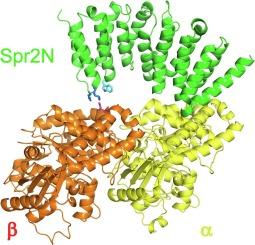
Abstract Image

负端靶向蛋白 Spiral2 与微管结合的结构分析
微管(MT)是一种动态细胞骨架聚合物,在决定细胞极性和形状方面起着至关重要的作用。在植物细胞中,顶体 MT 定位于细胞表面,被称为皮层 MT。皮层 MT 在细胞皮层成核,并从成核点分离。释放出来的MT丝进行踩踏运动,MT的正端聚合,负端解聚。负端靶向蛋白(-TIPs)包括螺旋2(Spiral2),它能调节顶体MT的负端动态。Spiral2 在 MT 负端自主聚集并抑制纤维收缩,但 Spiral2 特异性识别 MT 负端的机制仍不清楚。在这里,我们描述了 Spiral2 N 端 MT 结合结构域的晶体结构。该结构域的结构特性与肿瘤过表达基因(TOG)结构域的 HEAT 重复结构相似,但 HEAT 重复的数量不同,而且构象呈高度弧形。凝胶过滤和共沉淀分析表明,该结构域优先与 MT 细丝结合,而不是与微管蛋白二聚体结合,而且 Spiral2 通过基本表面与微管蛋白结合的模式与 TOG 结构域类似。我们构建了一个Spiral2-微管蛋白复合物的硅学模型,以确定可能识别微管蛋白的残基。突变分析表明,模型中推断出的关键残基参与了微管识别,并为末端靶向蛋白稳定MT末端的机制提供了启示。
本文章由计算机程序翻译,如有差异,请以英文原文为准。
求助全文
约1分钟内获得全文 求助全文
来源期刊
CiteScore
10.00
自引率
2.00%
发文量
151
审稿时长
44 days
期刊介绍: BBA Molecular Cell Research focuses on understanding the mechanisms of cellular processes at the molecular level. These include aspects of cellular signaling, signal transduction, cell cycle, apoptosis, intracellular trafficking, secretory and endocytic pathways, biogenesis of cell organelles, cytoskeletal structures, cellular interactions, cell/tissue differentiation and cellular enzymology. Also included are studies at the interface between Cell Biology and Biophysics which apply for example novel imaging methods for characterizing cellular processes.

×
引用
GB/T 7714-2015
复制
MLA
复制
APA
复制
导出至
BibTeX EndNote RefMan NoteFirst NoteExpress
×
提示
您的信息不完整,为了账户安全,请先补充。
现在去补充
×
提示
您因"违规操作"
具体请查看互助需知
我知道了
×
提示
确定
请完成安全验证×
copy
已复制链接
快去分享给好友吧!
我知道了
右上角分享
点击右上角分享
0
联系我们:info@booksci.cn Book学术提供免费学术资源搜索服务,方便国内外学者检索中英文文献。致力于提供最便捷和优质的服务体验。 Copyright © 2023 布克学术 All rights reserved.
京ICP备2023020795号-1
ghs 京公网安备 11010802042870号
Book学术文献互助
Book学术文献互助群
群 号:481959085
Book学术官方微信