{"title":"Convolutional Neural Network-Machine Learning Model: Hybrid Model for Meningioma Tumour and Healthy Brain Classification.","authors":"Simona Moldovanu, Gigi Tăbăcaru, Marian Barbu","doi":"10.3390/jimaging10090235","DOIUrl":null,"url":null,"abstract":"<p><p>This paper presents a hybrid study of convolutional neural networks (CNNs), machine learning (ML), and transfer learning (TL) in the context of brain magnetic resonance imaging (MRI). The anatomy of the brain is very complex; inside the skull, a brain tumour can form in any part. With MRI technology, cross-sectional images are generated, and radiologists can detect the abnormalities. When the size of the tumour is very small, it is undetectable to the human visual system, necessitating alternative analysis using AI tools. As is widely known, CNNs explore the structure of an image and provide features on the SoftMax fully connected (SFC) layer, and the classification of the items that belong to the input classes is established. Two comparison studies for the classification of meningioma tumours and healthy brains are presented in this paper: (i) classifying MRI images using an original CNN and two pre-trained CNNs, DenseNet169 and EfficientNetV2B0; (ii) determining which CNN and ML combination yields the most accurate classification when SoftMax is replaced with three ML models; in this context, Random Forest (RF), K-Nearest Neighbors (KNN), and Support Vector Machine (SVM) were proposed. In a binary classification of tumours and healthy brains, the EfficientNetB0-SVM combination shows an accuracy of 99.5% on the test dataset. A generalisation of the results was performed, and overfitting was prevented by using the bagging ensemble method.</p>","PeriodicalId":37035,"journal":{"name":"Journal of Imaging","volume":"10 9","pages":""},"PeriodicalIF":2.7000,"publicationDate":"2024-09-20","publicationTypes":"Journal Article","fieldsOfStudy":null,"isOpenAccess":false,"openAccessPdf":"https://www.ncbi.nlm.nih.gov/pmc/articles/PMC11433632/pdf/","citationCount":"0","resultStr":null,"platform":"Semanticscholar","paperid":null,"PeriodicalName":"Journal of Imaging","FirstCategoryId":"1085","ListUrlMain":"https://doi.org/10.3390/jimaging10090235","RegionNum":0,"RegionCategory":null,"ArticlePicture":[],"TitleCN":null,"AbstractTextCN":null,"PMCID":null,"EPubDate":"","PubModel":"","JCR":"Q3","JCRName":"IMAGING SCIENCE & PHOTOGRAPHIC TECHNOLOGY","Score":null,"Total":0}
引用次数: 0
Abstract
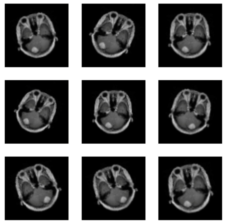
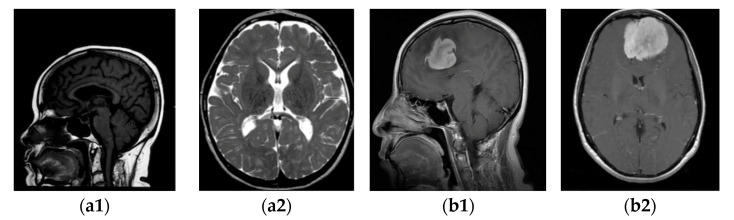
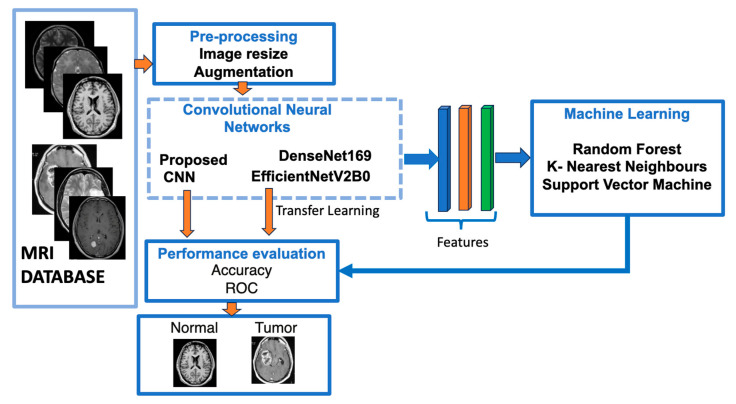
This paper presents a hybrid study of convolutional neural networks (CNNs), machine learning (ML), and transfer learning (TL) in the context of brain magnetic resonance imaging (MRI). The anatomy of the brain is very complex; inside the skull, a brain tumour can form in any part. With MRI technology, cross-sectional images are generated, and radiologists can detect the abnormalities. When the size of the tumour is very small, it is undetectable to the human visual system, necessitating alternative analysis using AI tools. As is widely known, CNNs explore the structure of an image and provide features on the SoftMax fully connected (SFC) layer, and the classification of the items that belong to the input classes is established. Two comparison studies for the classification of meningioma tumours and healthy brains are presented in this paper: (i) classifying MRI images using an original CNN and two pre-trained CNNs, DenseNet169 and EfficientNetV2B0; (ii) determining which CNN and ML combination yields the most accurate classification when SoftMax is replaced with three ML models; in this context, Random Forest (RF), K-Nearest Neighbors (KNN), and Support Vector Machine (SVM) were proposed. In a binary classification of tumours and healthy brains, the EfficientNetB0-SVM combination shows an accuracy of 99.5% on the test dataset. A generalisation of the results was performed, and overfitting was prevented by using the bagging ensemble method.

 求助内容:
求助内容: 应助结果提醒方式:


