MRI textural plasticity in limbic gray matter associated with clinical response to electroconvulsive therapy for psychosis

IF 9.6 1区 医学 Q1 BIOCHEMISTRY & MOLECULAR BIOLOGY
Eugenie Choe, Minah Kim, Sunah Choi, Harin Oh, Moonyoung Jang, Sunghyun Park, Jun Soo Kwon
{"title":"MRI textural plasticity in limbic gray matter associated with clinical response to electroconvulsive therapy for psychosis","authors":"Eugenie Choe, Minah Kim, Sunah Choi, Harin Oh, Moonyoung Jang, Sunghyun Park, Jun Soo Kwon","doi":"10.1038/s41380-024-02755-7","DOIUrl":null,"url":null,"abstract":"<p>Electroconvulsive therapy (ECT) is effective against treatment-resistant psychosis, but its mechanisms remain unclear. Conventional volumetry studies have revealed plasticity in limbic structures following ECT but with inconsistent clinical relevance, as they potentially overlook subtle histological alterations. Our study analyzed microstructural changes in limbic structures after ECT using MRI texture analysis and demonstrated a correlation with clinical response. 36 schizophrenia or schizoaffective patients treated with ECT and medication, 27 patients treated with medication only, and 70 healthy controls (HCs) were included in this study. Structural MRI data were acquired before and after ECT for the ECT group and at equivalent intervals for the medication-only group. The gray matter volume and MRI texture, calculated from the gray level size zone matrix (GLSZM), were extracted from limbic structures. After normalizing texture features to HC data, group-time interactions were estimated with repeated-measures mixed models. Repeated-measures correlations between clinical variables and texture were analyzed. Volumetric group-time interactions were observed in seven of fourteen limbic structures. Group-time interactions of the normalized GLSZM large area emphasis of the left hippocampus and the right amygdala reached statistical significance. Changes in these texture features were correlated with changes in psychotic symptoms in the ECT group but not in the medication-only group. These findings provide in vivo evidence that microstructural changes in key limbic structures, hypothetically reflected by MRI texture, are associated with clinical response to ECT for psychosis. These findings support the neuroplasticity hypothesis of ECT and highlight the hippocampus and amygdala as potential targets for neuromodulation in psychosis.</p>","PeriodicalId":19008,"journal":{"name":"Molecular Psychiatry","volume":"26 1","pages":""},"PeriodicalIF":9.6000,"publicationDate":"2024-09-26","publicationTypes":"Journal Article","fieldsOfStudy":null,"isOpenAccess":false,"openAccessPdf":"","citationCount":"0","resultStr":null,"platform":"Semanticscholar","paperid":null,"PeriodicalName":"Molecular Psychiatry","FirstCategoryId":"3","ListUrlMain":"https://doi.org/10.1038/s41380-024-02755-7","RegionNum":1,"RegionCategory":"医学","ArticlePicture":[],"TitleCN":null,"AbstractTextCN":null,"PMCID":null,"EPubDate":"","PubModel":"","JCR":"Q1","JCRName":"BIOCHEMISTRY & MOLECULAR BIOLOGY","Score":null,"Total":0}
引用次数: 0

Abstract

Electroconvulsive therapy (ECT) is effective against treatment-resistant psychosis, but its mechanisms remain unclear. Conventional volumetry studies have revealed plasticity in limbic structures following ECT but with inconsistent clinical relevance, as they potentially overlook subtle histological alterations. Our study analyzed microstructural changes in limbic structures after ECT using MRI texture analysis and demonstrated a correlation with clinical response. 36 schizophrenia or schizoaffective patients treated with ECT and medication, 27 patients treated with medication only, and 70 healthy controls (HCs) were included in this study. Structural MRI data were acquired before and after ECT for the ECT group and at equivalent intervals for the medication-only group. The gray matter volume and MRI texture, calculated from the gray level size zone matrix (GLSZM), were extracted from limbic structures. After normalizing texture features to HC data, group-time interactions were estimated with repeated-measures mixed models. Repeated-measures correlations between clinical variables and texture were analyzed. Volumetric group-time interactions were observed in seven of fourteen limbic structures. Group-time interactions of the normalized GLSZM large area emphasis of the left hippocampus and the right amygdala reached statistical significance. Changes in these texture features were correlated with changes in psychotic symptoms in the ECT group but not in the medication-only group. These findings provide in vivo evidence that microstructural changes in key limbic structures, hypothetically reflected by MRI texture, are associated with clinical response to ECT for psychosis. These findings support the neuroplasticity hypothesis of ECT and highlight the hippocampus and amygdala as potential targets for neuromodulation in psychosis.

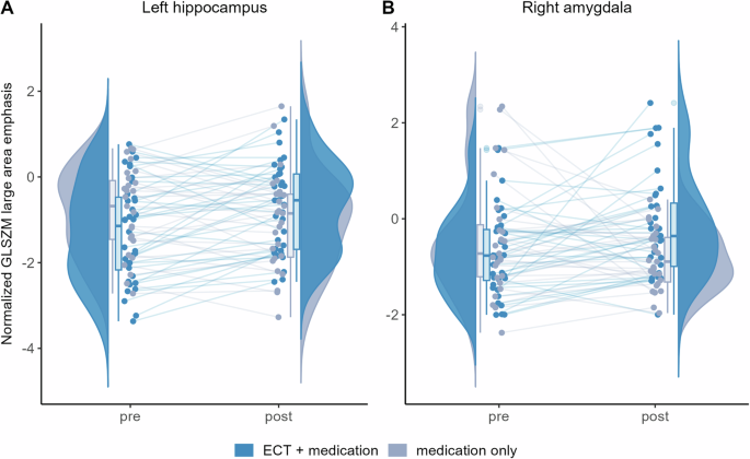
Abstract Image

边缘灰质的磁共振成像纹理可塑性与电休克疗法治疗精神病的临床反应有关
电休克疗法(ECT)能有效治疗耐药精神病,但其机制仍不清楚。传统的容积测量研究揭示了电休克疗法后边缘结构的可塑性,但其临床意义并不一致,因为它们可能会忽略细微的组织学改变。我们的研究使用核磁共振成像纹理分析法分析了 ECT 后边缘结构的微观结构变化,并证明了其与临床反应的相关性。本研究共纳入了 36 名接受电痉挛疗法和药物治疗的精神分裂症或分裂情感障碍患者、27 名仅接受药物治疗的患者和 70 名健康对照组(HCs)。电痉挛治疗组在电痉挛治疗前后采集了结构磁共振成像数据,纯药物治疗组在相同时间间隔采集了结构磁共振成像数据。从边缘结构中提取了灰质体积和磁共振成像纹理(由灰度大小区矩阵(GLSZM)计算得出)。将纹理特征归一化为 HC 数据后,使用重复测量混合模型估计组间交互作用。分析了临床变量与纹理之间的重复测量相关性。在 14 个边缘结构中,有 7 个观察到了体积组-时间交互作用。左侧海马和右侧杏仁核的归一化 GLSZM 大面积重点的组时交互作用达到了统计学意义。这些纹理特征的变化与电痉挛疗法组精神病症状的变化相关,但与单纯药物治疗组无关。这些研究结果提供了活体证据,证明关键边缘结构的微观结构变化与治疗精神病的电痉挛疗法的临床反应有关,而这些变化正是核磁共振成像纹理所假定反映的。这些发现支持电痉挛疗法的神经可塑性假说,并强调海马和杏仁核是神经调节治疗精神病的潜在目标。
本文章由计算机程序翻译,如有差异,请以英文原文为准。
求助全文
约1分钟内获得全文 求助全文
来源期刊
Molecular Psychiatry
Molecular Psychiatry 医学-精神病学
CiteScore
20.50
自引率
4.50%
发文量
459
审稿时长
4-8 weeks
期刊介绍: Molecular Psychiatry focuses on publishing research that aims to uncover the biological mechanisms behind psychiatric disorders and their treatment. The journal emphasizes studies that bridge pre-clinical and clinical research, covering cellular, molecular, integrative, clinical, imaging, and psychopharmacology levels.
×
引用
GB/T 7714-2015
复制
MLA
复制
APA
复制
导出至
BibTeX EndNote RefMan NoteFirst NoteExpress
×
提示
您的信息不完整,为了账户安全,请先补充。
现在去补充
×
提示
您因"违规操作"
具体请查看互助需知
我知道了
×
提示
确定
请完成安全验证×
copy
已复制链接
快去分享给好友吧!
我知道了
右上角分享
点击右上角分享
0
联系我们:info@booksci.cn Book学术提供免费学术资源搜索服务,方便国内外学者检索中英文文献。致力于提供最便捷和优质的服务体验。 Copyright © 2023 布克学术 All rights reserved.
京ICP备2023020795号-1
ghs 京公网安备 11010802042870号
Book学术文献互助
Book学术文献互助群
群 号:481959085
Book学术官方微信