Fetal body organ T2* relaxometry at low field strength (FOREST)

IF 10.7 1区 医学 Q1 COMPUTER SCIENCE, ARTIFICIAL INTELLIGENCE
Kelly Payette , Alena U. Uus , Jordina Aviles Verdera , Megan Hall , Alexia Egloff , Maria Deprez , Raphaël Tomi-Tricot , Joseph V. Hajnal , Mary A. Rutherford , Lisa Story , Jana Hutter
{"title":"Fetal body organ T2* relaxometry at low field strength (FOREST)","authors":"Kelly Payette ,&nbsp;Alena U. Uus ,&nbsp;Jordina Aviles Verdera ,&nbsp;Megan Hall ,&nbsp;Alexia Egloff ,&nbsp;Maria Deprez ,&nbsp;Raphaël Tomi-Tricot ,&nbsp;Joseph V. Hajnal ,&nbsp;Mary A. Rutherford ,&nbsp;Lisa Story ,&nbsp;Jana Hutter","doi":"10.1016/j.media.2024.103352","DOIUrl":null,"url":null,"abstract":"<div><div>Fetal Magnetic Resonance Imaging (MRI) at low field strengths is an exciting new field in both clinical and research settings. Clinical low field (0.55T) scanners are beneficial for fetal imaging due to their reduced susceptibility-induced artifacts, increased T2* values, and wider bore (widening access for the increasingly obese pregnant population). However, the lack of standard automated image processing tools such as segmentation and reconstruction hampers wider clinical use. In this study, we present the Fetal body Organ T2* RElaxometry at low field STrength (FOREST) pipeline that analyzes ten major fetal body organs. Dynamic multi-echo multi-gradient sequences were acquired and automatically reoriented to a standard plane, reconstructed into a high-resolution volume using deformable slice-to-volume reconstruction, and then automatically segmented into ten major fetal organs. We extensively validated FOREST using an inter-rater quality analysis. We then present fetal T2* body organ growth curves made from 100 control subjects from a wide gestational age range (17–40 gestational weeks) in order to investigate the relationship of T2* with gestational age. The T2* values for all organs except the stomach and spleen were found to have a relationship with gestational age (p<span><math><mo>&lt;</mo></math></span>0.05). FOREST is robust to fetal motion, and can be used for both normal and fetuses with pathologies. Low field fetal MRI can be used to perform advanced MRI analysis, and is a viable option for clinical scanning.</div></div>","PeriodicalId":18328,"journal":{"name":"Medical image analysis","volume":"99 ","pages":"Article 103352"},"PeriodicalIF":10.7000,"publicationDate":"2024-09-19","publicationTypes":"Journal Article","fieldsOfStudy":null,"isOpenAccess":false,"openAccessPdf":"","citationCount":"0","resultStr":null,"platform":"Semanticscholar","paperid":null,"PeriodicalName":"Medical image analysis","FirstCategoryId":"5","ListUrlMain":"https://www.sciencedirect.com/science/article/pii/S1361841524002779","RegionNum":1,"RegionCategory":"医学","ArticlePicture":[],"TitleCN":null,"AbstractTextCN":null,"PMCID":null,"EPubDate":"","PubModel":"","JCR":"Q1","JCRName":"COMPUTER SCIENCE, ARTIFICIAL INTELLIGENCE","Score":null,"Total":0}
引用次数: 0

Abstract

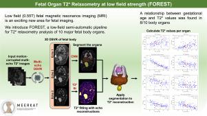
Fetal Magnetic Resonance Imaging (MRI) at low field strengths is an exciting new field in both clinical and research settings. Clinical low field (0.55T) scanners are beneficial for fetal imaging due to their reduced susceptibility-induced artifacts, increased T2* values, and wider bore (widening access for the increasingly obese pregnant population). However, the lack of standard automated image processing tools such as segmentation and reconstruction hampers wider clinical use. In this study, we present the Fetal body Organ T2* RElaxometry at low field STrength (FOREST) pipeline that analyzes ten major fetal body organs. Dynamic multi-echo multi-gradient sequences were acquired and automatically reoriented to a standard plane, reconstructed into a high-resolution volume using deformable slice-to-volume reconstruction, and then automatically segmented into ten major fetal organs. We extensively validated FOREST using an inter-rater quality analysis. We then present fetal T2* body organ growth curves made from 100 control subjects from a wide gestational age range (17–40 gestational weeks) in order to investigate the relationship of T2* with gestational age. The T2* values for all organs except the stomach and spleen were found to have a relationship with gestational age (p<0.05). FOREST is robust to fetal motion, and can be used for both normal and fetuses with pathologies. Low field fetal MRI can be used to perform advanced MRI analysis, and is a viable option for clinical scanning.
Abstract Image

低场强胎儿身体器官 T2* 弛豫测量(FOREST)
低磁场强度胎儿磁共振成像(MRI)在临床和研究领域都是一个令人兴奋的新领域。临床用低磁场(0.55T)扫描仪由于减少了由易感性引起的伪影、提高了 T2* 值和更宽的孔径(为日益肥胖的孕妇提供了更多的检查机会)而有利于胎儿成像。然而,由于缺乏标准的自动图像处理工具(如分割和重建),阻碍了其在临床上的广泛应用。在这项研究中,我们介绍了胎儿体内器官 T2* RElaxometry at low field STrength (FOREST) 管道,该管道可分析胎儿体内的十个主要器官。我们采集了动态多回波多梯度序列,并将其自动调整到标准平面,利用可变形切片-体积重建技术重建成高分辨率体积,然后自动分割成十个主要胎儿器官。我们利用评分者之间的质量分析对 FOREST 进行了广泛验证。然后,我们展示了 100 名胎龄范围较宽(17-40 孕周)的对照受试者的胎儿 T2* 身体器官生长曲线,以研究 T2* 与胎龄的关系。结果发现,除胃和脾脏外,所有器官的 T2* 值都与胎龄有关(p<0.05)。FOREST对胎儿的运动很敏感,既可用于正常胎儿,也可用于有病变的胎儿。低场胎儿磁共振成像可用于进行高级磁共振成像分析,是临床扫描的可行选择。
本文章由计算机程序翻译,如有差异,请以英文原文为准。
求助全文
约1分钟内获得全文 求助全文
来源期刊
Medical image analysis
Medical image analysis 工程技术-工程:生物医学
CiteScore
22.10
自引率
6.40%
发文量
309
审稿时长
6.6 months
期刊介绍: Medical Image Analysis serves as a platform for sharing new research findings in the realm of medical and biological image analysis, with a focus on applications of computer vision, virtual reality, and robotics to biomedical imaging challenges. The journal prioritizes the publication of high-quality, original papers contributing to the fundamental science of processing, analyzing, and utilizing medical and biological images. It welcomes approaches utilizing biomedical image datasets across all spatial scales, from molecular/cellular imaging to tissue/organ imaging.
×
引用
GB/T 7714-2015
复制
MLA
复制
APA
复制
导出至
BibTeX EndNote RefMan NoteFirst NoteExpress
×
提示
您的信息不完整,为了账户安全,请先补充。
现在去补充
×
提示
您因"违规操作"
具体请查看互助需知
我知道了
×
提示
确定
请完成安全验证×
copy
已复制链接
快去分享给好友吧!
我知道了
右上角分享
点击右上角分享
0
联系我们:info@booksci.cn Book学术提供免费学术资源搜索服务,方便国内外学者检索中英文文献。致力于提供最便捷和优质的服务体验。 Copyright © 2023 布克学术 All rights reserved.
京ICP备2023020795号-1
ghs 京公网安备 11010802042870号
Book学术文献互助
Book学术文献互助群
群 号:481959085
Book学术官方微信