Evaluation of the geometric and dosimetric accuracies of deformable image registration of targets and critical organs in prostate CBCT-guided adaptive radiotherapy
Abstract
Purpose
Kilovoltage cone beam computed tomography (kVCBCT)-guided adaptive radiation therapy (ART) uses daily deformed CT (dCT), which is generated automatically through deformable registration methods. These registration methods may perform poorly in reproducing volumes of the target organ, rectum, and bladder during treatment. We analyzed the registration errors between the daily kVCBCTs and corresponding dCTs for these organs using the default optical flow algorithm and two registration procedures. We validated the effectiveness of these registration methods in replicating the geometry for dose calculation on kVCBCT for ART.
Methods
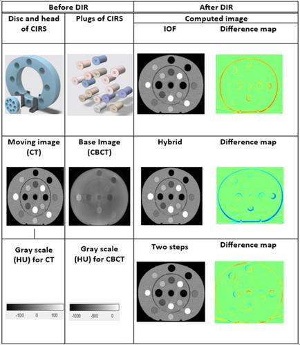
We evaluated three deformable image registration (DIR) methods to assess their registration accuracy and dose calculation effeciency in mapping target and critical organs. The DIR methods include (1) default intensity-based deformable registration, (2) hybrid deformable registration, and (3) a two-step deformable registration process. Each technique was applied to a computerized imaging reference system (CIRS) phantom (Model 062 M) and to five patients who received volumetric modulated arc therapy to the prostate. Registration accuracy was assessed using the 95% Hausdorff distance (HD95) and Dice similarity coefficient (DSC), and each method was compared with the intensity-based registration method. The improvement in the dCT image quality of the CIRS phantom and five patients was assessed by comparing dCT with kVCBCT. Image quality quantitative metrics for the phantom included the signal-to-noise ratio (SNR), uniformity, and contrast-to-noise ratio (CNR), whereas those for the patients included the mean absolute error (MAE), mean error, peak signal-to-noise ratio (PSNR), and structural similarity index measure (SSIM). To determine dose metric differences, we used a dose-volume histogram (DVH) and 3.0%/0.3 mm gamma analysis to compare planning computed tomography (pCT) and kVCBCT recalculations with restimulated CT images used as a reference.
Results
The dCT images generated by the hybrid (dCTH) and two-step (dCTC) registration methods resulted in significant improvements compared to kVCBCT in the phantom model. Specifically, the SNR improved by 107% and 107.2%, the uniformity improved by 90% and 75%, and the CNR improved by 212.2% and 225.6 for dCTH and dCTC methods, respectively. For the patient images, the MAEs improved by 98% and 94%, the PSNRs improved by 16.3% and 22.9%, and the SSIMs improved by 1% and 1% in the dCTH and dCTC methods, respectively. For the geometric evaluation, only the two-step registration method improved registration accuracy. The dCTH method yielded an average HD95 of 12 mm and average DSC of 0.73, whereas dCTC yielded an average HD95 of 2.9 mm and average DSC of 0.902. The DVH showed that the dCTC-based dose calculations differed by <2% from the expected results for treatment targets and volumes of organs at risk. Additionally, gamma indices for dCTC-based treatment plans were >95% at all points, whereas they were <95% for kVCBCT-based treatment plans.
Conclusion
The two-step registration method outperforms the intensity-based and hybrid registration methods. While the hybrid and two-step-based methods improved the image quality of kVCBCT in a linear accelerator, only the two-step method improved the registration accuracy of the corresponding structures among the pCT and kVCBCT datasets. A two-step registration process is recommended for applying kVCBCT to ART, which achieves better registration accuracy for local and global image structures. This method appears to be beneficial for radiotherapy dose calculation in patients with pelvic cancer.


 求助内容:
求助内容: 应助结果提醒方式:


