Xia-Qing Sheng, Zi-Han Peng, Nan-Fang Pan, You-Jin Zhao, Quan Gong, Yue-Ming Song, Qi-Yong Gong, Hao Liu, Yang Meng
{"title":"Novel MRI signs of the atlantodental space in patients with atlantoaxial dislocation","authors":"Xia-Qing Sheng, Zi-Han Peng, Nan-Fang Pan, You-Jin Zhao, Quan Gong, Yue-Ming Song, Qi-Yong Gong, Hao Liu, Yang Meng","doi":"10.1007/s00586-024-08498-x","DOIUrl":null,"url":null,"abstract":"<h3 data-test=\"abstract-sub-heading\">Objectives</h3><p>The type of atlantodental space tissue in patients with atlantoaxial dislocation (AAD) can help doctors understand the possibility of reduction before surgery. However, relevant research on this topic is lacking. In this study, we aimed to summarise the atlantodental space classification of patients with AAD using magnetic resonance imaging (MRI) and explore their clinical characteristics.</p><h3 data-test=\"abstract-sub-heading\">Materials and methods</h3><p>Preoperative 3T cervical MR images of patients who underwent posterior reduction and fixation surgery for non-traumatic AAD between 1 September 2012 and 31 July 2023 were collected. Two radiologists read and recorded the MRI results based on the standard protocol. The kappa value was used to evaluate intra- and inter-observer agreements. The patient’s age, sex, body mass index, clinical symptoms, Japanese Orthopaedic Association (JOA) score, and visual analogue scale information were obtained from medical records.</p><h3 data-test=\"abstract-sub-heading\">Results</h3><p>A total of 135 patients with AAD (mean age, 51.3 ± 14.0 years, 52 men) were included in the analysis. The inter-observer agreement between the two readers was 0.818 (<i>P</i> < 0.0001). The intra-observer consistencies were 0.882 (<i>P</i> < 0.0001) and 0.896 (<i>P</i> < 0.0001). Patients with inflexible tissue signs exhibit more irreducible in hyperextension position, and their range of motion of ADI is smaller. These patients were older and had a higher incidence of abnormal spinal cord signals and JOA scores.</p><h3 data-test=\"abstract-sub-heading\">Conclusions</h3><p>Novel MRI signs exhibited high inter- and intra-observer consistency and were associated with patient age, abnormal spinal cord signals, reducibility, range of motion of ADI, and symptoms.</p>","PeriodicalId":12323,"journal":{"name":"European Spine Journal","volume":null,"pages":null},"PeriodicalIF":2.6000,"publicationDate":"2024-09-19","publicationTypes":"Journal Article","fieldsOfStudy":null,"isOpenAccess":false,"openAccessPdf":"","citationCount":"0","resultStr":null,"platform":"Semanticscholar","paperid":null,"PeriodicalName":"European Spine Journal","FirstCategoryId":"3","ListUrlMain":"https://doi.org/10.1007/s00586-024-08498-x","RegionNum":3,"RegionCategory":"医学","ArticlePicture":[],"TitleCN":null,"AbstractTextCN":null,"PMCID":null,"EPubDate":"","PubModel":"","JCR":"Q2","JCRName":"CLINICAL NEUROLOGY","Score":null,"Total":0}
引用次数: 0
Abstract
Objectives
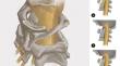
The type of atlantodental space tissue in patients with atlantoaxial dislocation (AAD) can help doctors understand the possibility of reduction before surgery. However, relevant research on this topic is lacking. In this study, we aimed to summarise the atlantodental space classification of patients with AAD using magnetic resonance imaging (MRI) and explore their clinical characteristics.
Materials and methods
Preoperative 3T cervical MR images of patients who underwent posterior reduction and fixation surgery for non-traumatic AAD between 1 September 2012 and 31 July 2023 were collected. Two radiologists read and recorded the MRI results based on the standard protocol. The kappa value was used to evaluate intra- and inter-observer agreements. The patient’s age, sex, body mass index, clinical symptoms, Japanese Orthopaedic Association (JOA) score, and visual analogue scale information were obtained from medical records.
Results
A total of 135 patients with AAD (mean age, 51.3 ± 14.0 years, 52 men) were included in the analysis. The inter-observer agreement between the two readers was 0.818 (P < 0.0001). The intra-observer consistencies were 0.882 (P < 0.0001) and 0.896 (P < 0.0001). Patients with inflexible tissue signs exhibit more irreducible in hyperextension position, and their range of motion of ADI is smaller. These patients were older and had a higher incidence of abnormal spinal cord signals and JOA scores.
Conclusions
Novel MRI signs exhibited high inter- and intra-observer consistency and were associated with patient age, abnormal spinal cord signals, reducibility, range of motion of ADI, and symptoms.
期刊介绍:
"European Spine Journal" is a publication founded in response to the increasing trend toward specialization in spinal surgery and spinal pathology in general. The Journal is devoted to all spine related disciplines, including functional and surgical anatomy of the spine, biomechanics and pathophysiology, diagnostic procedures, and neurology, surgery and outcomes. The aim of "European Spine Journal" is to support the further development of highly innovative spine treatments including but not restricted to surgery and to provide an integrated and balanced view of diagnostic, research and treatment procedures as well as outcomes that will enhance effective collaboration among specialists worldwide. The “European Spine Journal” also participates in education by means of videos, interactive meetings and the endorsement of educative efforts.
Official publication of EUROSPINE, The Spine Society of Europe

 求助内容:
求助内容: 应助结果提醒方式:


