A microscopic oxygen transport model for ultra-high dose rate radiotherapy in vivo: The impact of physiological conditions on FLASH effect

IF 3.2 2区 医学 Q1 RADIOLOGY, NUCLEAR MEDICINE & MEDICAL IMAGING
Medical physics Pub Date : 2024-09-16 DOI:10.1002/mp.17398
Lixiang Guo, Paul M. Medin, Ken Kang-Hsin Wang
{"title":"A microscopic oxygen transport model for ultra-high dose rate radiotherapy in vivo: The impact of physiological conditions on FLASH effect","authors":"Lixiang Guo,&nbsp;Paul M. Medin,&nbsp;Ken Kang-Hsin Wang","doi":"10.1002/mp.17398","DOIUrl":null,"url":null,"abstract":"<div>\n \n \n <section>\n \n <h3> Background</h3>\n \n <p>Ultra-high dose rate irradiation (≥40 Gy/s, FLASH) has been shown to reduce normal tissue toxicity, while maintaining tumor control compared to conventional dose-rate radiotherapy. The radiolytic oxygen (O<sub>2</sub>) depletion (ROD) resulting from FLASH has been proposed to explain the normal tissue protection effect; however, in vivo experiments have not confirmed that FLASH induced global tissue hypoxia. Nonetheless, the experiments reported are based on volume-averaged measurement, which have inherent limitations in detecting microscopic phenomena, including the potential preservation of stem cells niches due to local FLASH-induced O<sub>2</sub> depletion. Computational modeling offers a complementary approach to understand the ROD caused by FLASH at the microscopic level.</p>\n </section>\n \n <section>\n \n <h3> Purpose</h3>\n \n <p>We developed a comprehensive model to describe the spatial and temporal dynamics of O<sub>2</sub> consumption and transport in response to irradiation in vivo. The change of oxygen enhancement ratio (OER) was used to quantify and investigate the FLASH effect as a function of physiological and radiation parameters at microscopic scale.</p>\n </section>\n \n <section>\n \n <h3> Methods</h3>\n \n <p>We considered time-dependent O<sub>2</sub> supply and consumption in a 3D cylindrical geometry, incorporating blood flow linking the O<sub>2</sub> concentration ([O<sub>2</sub>]) in the capillary to that within the tissue through the Hill equation, radial and axial diffusion of O<sub>2</sub>, metabolic and zero-order radiolytic O<sub>2</sub> consumption, and a pulsed radiation structure. Time-evolved distributions of [O<sub>2</sub>] were obtained by numerically solving perfusion-diffusion equations. The model enables the computation of dynamic O<sub>2</sub> distribution and the relative change of OER (<i>δ<sub>ROD</sub></i>) under various physiological and radiation conditions in vivo.</p>\n </section>\n \n <section>\n \n <h3> Results</h3>\n \n <p>Initial [O<sub>2</sub>] level and the subsequent changes during irradiation determined <i>δ<sub>ROD</sub></i> distribution, which strongly depends on physiological parameters, i.e., intercapillary spacing, ultimately determining the tissue area with enhanced radioresistance. We observed that the <i>δ<sub>ROD</sub></i>/FLASH effect is affected by and sensitive to the interplay effect among physiological and radiation parameters. It renders that the FLASH effect can be tissue environment dependent. The saturation of FLASH normal tissue protection upon dose and dose rate was shown. Beyond ∼60 Gy/s, no significant decrease in radiosensitivity within tissue region was observed. In turn, for a given dose rate, the change of radiosensitivity became saturated after a certain dose level. Pulse structures with the same dose and instantaneous dose rate but with different delivery times were shown to have distinguishable <i>δ<sub>ROD</sub></i> thus tissue sparing, suggesting the average dose rate could be a metric assessing the FLASH effect and demonstrating the capability of our model to support experimental findings.</p>\n </section>\n \n <section>\n \n <h3> Conclusion</h3>\n \n <p>On a macroscopic scale, the modeling results align with the experimental findings in terms of dose and dose rate thresholds, and it also indicates that pulse structure can vary the FLASH effect. At the microscopic level, this model enables us to examine the spatially resolved FLASH effect based on physiological and irradiation parameters. Our model thus provides a complementary approach to experimental methods for understanding the underlying mechanism of FLASH radiotherapy. Our results show that physiological conditions can potentially determine the FLASH efficacy in tissue protection. The FLASH effect may be observed under optimal combination of physiological parameters, not limited to radiation conditions alone.</p>\n </section>\n </div>","PeriodicalId":18384,"journal":{"name":"Medical physics","volume":"51 11","pages":"8623-8637"},"PeriodicalIF":3.2000,"publicationDate":"2024-09-16","publicationTypes":"Journal Article","fieldsOfStudy":null,"isOpenAccess":false,"openAccessPdf":"https://onlinelibrary.wiley.com/doi/epdf/10.1002/mp.17398","citationCount":"0","resultStr":null,"platform":"Semanticscholar","paperid":null,"PeriodicalName":"Medical physics","FirstCategoryId":"3","ListUrlMain":"https://onlinelibrary.wiley.com/doi/10.1002/mp.17398","RegionNum":2,"RegionCategory":"医学","ArticlePicture":[],"TitleCN":null,"AbstractTextCN":null,"PMCID":null,"EPubDate":"","PubModel":"","JCR":"Q1","JCRName":"RADIOLOGY, NUCLEAR MEDICINE & MEDICAL IMAGING","Score":null,"Total":0}
引用次数: 0

Abstract

Background

Ultra-high dose rate irradiation (≥40 Gy/s, FLASH) has been shown to reduce normal tissue toxicity, while maintaining tumor control compared to conventional dose-rate radiotherapy. The radiolytic oxygen (O2) depletion (ROD) resulting from FLASH has been proposed to explain the normal tissue protection effect; however, in vivo experiments have not confirmed that FLASH induced global tissue hypoxia. Nonetheless, the experiments reported are based on volume-averaged measurement, which have inherent limitations in detecting microscopic phenomena, including the potential preservation of stem cells niches due to local FLASH-induced O2 depletion. Computational modeling offers a complementary approach to understand the ROD caused by FLASH at the microscopic level.

Purpose

We developed a comprehensive model to describe the spatial and temporal dynamics of O2 consumption and transport in response to irradiation in vivo. The change of oxygen enhancement ratio (OER) was used to quantify and investigate the FLASH effect as a function of physiological and radiation parameters at microscopic scale.

Methods

We considered time-dependent O2 supply and consumption in a 3D cylindrical geometry, incorporating blood flow linking the O2 concentration ([O2]) in the capillary to that within the tissue through the Hill equation, radial and axial diffusion of O2, metabolic and zero-order radiolytic O2 consumption, and a pulsed radiation structure. Time-evolved distributions of [O2] were obtained by numerically solving perfusion-diffusion equations. The model enables the computation of dynamic O2 distribution and the relative change of OER (δROD) under various physiological and radiation conditions in vivo.

Results

Initial [O2] level and the subsequent changes during irradiation determined δROD distribution, which strongly depends on physiological parameters, i.e., intercapillary spacing, ultimately determining the tissue area with enhanced radioresistance. We observed that the δROD/FLASH effect is affected by and sensitive to the interplay effect among physiological and radiation parameters. It renders that the FLASH effect can be tissue environment dependent. The saturation of FLASH normal tissue protection upon dose and dose rate was shown. Beyond ∼60 Gy/s, no significant decrease in radiosensitivity within tissue region was observed. In turn, for a given dose rate, the change of radiosensitivity became saturated after a certain dose level. Pulse structures with the same dose and instantaneous dose rate but with different delivery times were shown to have distinguishable δROD thus tissue sparing, suggesting the average dose rate could be a metric assessing the FLASH effect and demonstrating the capability of our model to support experimental findings.

Conclusion

On a macroscopic scale, the modeling results align with the experimental findings in terms of dose and dose rate thresholds, and it also indicates that pulse structure can vary the FLASH effect. At the microscopic level, this model enables us to examine the spatially resolved FLASH effect based on physiological and irradiation parameters. Our model thus provides a complementary approach to experimental methods for understanding the underlying mechanism of FLASH radiotherapy. Our results show that physiological conditions can potentially determine the FLASH efficacy in tissue protection. The FLASH effect may be observed under optimal combination of physiological parameters, not limited to radiation conditions alone.
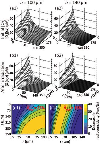
Abstract Image

体内超高剂量率放射治疗的微观氧传输模型:生理条件对 FLASH 效应的影响
背景超高剂量率照射(≥40 Gy/s,FLASH)与传统剂量率放疗相比,可降低正常组织毒性,同时保持肿瘤控制。有人提出,FLASH 导致的放射性氧(O2)耗竭(ROD)可解释正常组织保护效应;然而,体内实验并未证实 FLASH 会诱发整体组织缺氧。然而,所报道的实验都是基于体积平均测量,在检测微观现象方面存在固有的局限性,包括FLASH诱导的局部氧气耗竭可能导致干细胞龛位的保存。我们建立了一个全面的模型来描述体内辐照时氧气消耗和运输的时空动态。方法我们考虑了三维圆柱几何中随时间变化的氧气供应和消耗,通过希尔方程将毛细血管中的氧气浓度([O2])与组织内的氧气浓度([O2])联系起来的血流、氧气的径向和轴向扩散、代谢和零阶放射性氧气消耗以及脉冲辐射结构。通过对灌注-扩散方程进行数值求解,获得了[O2]的时变分布。结果初始[O2]水平和随后在辐照过程中的变化决定了δROD的分布,而δROD的分布与生理参数(即毛细血管间距)密切相关,最终决定了放射抗性增强的组织区域。我们观察到,δROD/FLASH 效应受生理参数和辐射参数之间相互作用效应的影响,而且对这种相互作用效应非常敏感。因此,FLASH效应可能与组织环境有关。FLASH对正常组织的保护在剂量和剂量率的作用下达到饱和。超过 60 Gy/s,组织区域内的辐射敏感性没有明显下降。反过来,对于给定的剂量率,辐射敏感性的变化在一定剂量水平后趋于饱和。结果表明,具有相同剂量和瞬时剂量率但不同投射时间的脉冲结构具有可区分的δROD,从而具有组织疏通性,这表明平均剂量率可以作为评估 FLASH 效应的指标,并证明了我们的模型有能力支持实验结果。在微观层面,该模型使我们能够根据生理和辐照参数来研究空间分辨的 FLASH 效应。因此,我们的模型为了解 FLASH 放射治疗的基本机制提供了一种与实验方法互补的方法。我们的结果表明,生理条件有可能决定 FLASH 在组织保护方面的功效。在最佳生理参数组合下可以观察到FLASH效应,而不仅仅局限于辐射条件。
本文章由计算机程序翻译,如有差异,请以英文原文为准。
求助全文
约1分钟内获得全文 求助全文
来源期刊
Medical physics
Medical physics 医学-核医学
CiteScore
6.80
自引率
15.80%
发文量
660
审稿时长
1.7 months
期刊介绍: Medical Physics publishes original, high impact physics, imaging science, and engineering research that advances patient diagnosis and therapy through contributions in 1) Basic science developments with high potential for clinical translation 2) Clinical applications of cutting edge engineering and physics innovations 3) Broadly applicable and innovative clinical physics developments Medical Physics is a journal of global scope and reach. By publishing in Medical Physics your research will reach an international, multidisciplinary audience including practicing medical physicists as well as physics- and engineering based translational scientists. We work closely with authors of promising articles to improve their quality.
×
引用
GB/T 7714-2015
复制
MLA
复制
APA
复制
导出至
BibTeX EndNote RefMan NoteFirst NoteExpress
×
提示
您的信息不完整,为了账户安全,请先补充。
现在去补充
×
提示
您因"违规操作"
具体请查看互助需知
我知道了
×
提示
确定
请完成安全验证×
copy
已复制链接
快去分享给好友吧!
我知道了
右上角分享
点击右上角分享
0
联系我们:info@booksci.cn Book学术提供免费学术资源搜索服务,方便国内外学者检索中英文文献。致力于提供最便捷和优质的服务体验。 Copyright © 2023 布克学术 All rights reserved.
京ICP备2023020795号-1
ghs 京公网安备 11010802042870号
Book学术文献互助
Book学术文献互助群
群 号:481959085
Book学术官方微信