{"title":"Man with severe neck pain","authors":"Sho Yamaguchi MD, Yuya Kitai MD, Tetsuya Inoue MD, PhD","doi":"10.1002/emp2.13276","DOIUrl":null,"url":null,"abstract":"<p>A 53-year-old male presented with acute neck pain radiating to the occiput for 2 days. He had been playing golf daily before symptom onset. There was no history of recent upper respiratory infection. Examination revealed an axillary temperature of 37.4°C, with other vital signs normal. The patient was alert with no meningeal signs, and neck pain worsened with rotation. Neurological examination was normal, with no palpable lymphadenopathy, and the pharyngeal examination was normal. Computed tomography (CT) confirmed the diagnosis (Figure 1).</p><p>The authors declare no conflicts of interest.</p>","PeriodicalId":73967,"journal":{"name":"Journal of the American College of Emergency Physicians open","volume":null,"pages":null},"PeriodicalIF":1.6000,"publicationDate":"2024-09-17","publicationTypes":"Journal Article","fieldsOfStudy":null,"isOpenAccess":false,"openAccessPdf":"https://onlinelibrary.wiley.com/doi/epdf/10.1002/emp2.13276","citationCount":"0","resultStr":null,"platform":"Semanticscholar","paperid":null,"PeriodicalName":"Journal of the American College of Emergency Physicians open","FirstCategoryId":"1085","ListUrlMain":"https://onlinelibrary.wiley.com/doi/10.1002/emp2.13276","RegionNum":0,"RegionCategory":null,"ArticlePicture":[],"TitleCN":null,"AbstractTextCN":null,"PMCID":null,"EPubDate":"","PubModel":"","JCR":"Q2","JCRName":"EMERGENCY MEDICINE","Score":null,"Total":0}
引用次数: 0
Abstract
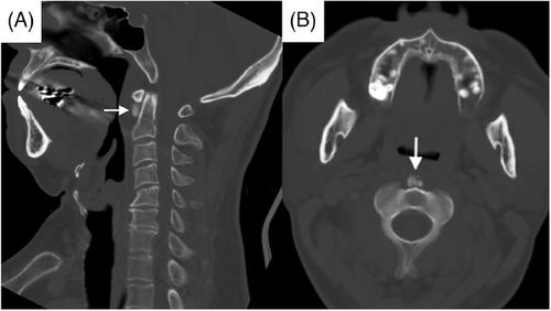
A 53-year-old male presented with acute neck pain radiating to the occiput for 2 days. He had been playing golf daily before symptom onset. There was no history of recent upper respiratory infection. Examination revealed an axillary temperature of 37.4°C, with other vital signs normal. The patient was alert with no meningeal signs, and neck pain worsened with rotation. Neurological examination was normal, with no palpable lymphadenopathy, and the pharyngeal examination was normal. Computed tomography (CT) confirmed the diagnosis (Figure 1).