{"title":"Evaluation of cerebellar aging in MRI images: Fractal analysis compared to Euclidean geometry-based morphometry","authors":"Nataliia Maryenko, Oleksandr Stepanenko","doi":"10.1016/j.metrad.2024.100101","DOIUrl":null,"url":null,"abstract":"<div><h3>Objectives</h3><p>This study aimed to identify age-related changes in the fractal dimensions of the cerebellum and compare the sensitivity of fractal analysis and conventional Euclidean geometry-based morphometry to cerebellar aging.</p></div><div><h3>Material and methods</h3><p>Two-dimensional T2-weighted magnetic resonance images from the brains of 100 conditionally healthy individuals (44 males and 56 females) aged 18–86 years were examined, with a focus on mid-sagittal sections of the cerebellar vermis. We determined ten parameters derived from Euclidean geometry (perimeter, area, and indices calculated from them), along with seven fractal dimension values derived from fractal geometry (the approximated fractal dimensions of the overall cerebellar tissue, white matter, overall cerebellar cortex and its granular and molecular layers, outer contour, and digital skeleton).</p></div><div><h3>Results</h3><p>Fractal dimensions demonstrated stronger correlation relationships with age compared to morphometric parameters derived from Euclidean geometry. The most pronounced age-related declines were observed in the approximated fractal dimensions of the cerebellar cortex and its layers, with decreases also noted in the fractal dimensions of the outer contour and digital cerebellar skeleton. Fractal dimension values did not significantly differ between males and females, while several Euclidean geometry-derived parameters showed sexual dimorphism. Although males demonstrated stronger relationships of some studied parameters with age, there was no statistically significant difference in the sex-related dynamics of aging.</p></div><div><h3>Conclusion</h3><p>The normal aging of the cerebellum involves not only absolute size alterations but also changes in the texture and spatial configuration of cerebellar tissue components, which can be quantitatively and objectively assessed by fractal analysis.</p></div>","PeriodicalId":100921,"journal":{"name":"Meta-Radiology","volume":"2 3","pages":"Article 100101"},"PeriodicalIF":0.0000,"publicationDate":"2024-09-01","publicationTypes":"Journal Article","fieldsOfStudy":null,"isOpenAccess":false,"openAccessPdf":"https://www.sciencedirect.com/science/article/pii/S2950162824000559/pdfft?md5=30f5636baa7a517496d299b30725e948&pid=1-s2.0-S2950162824000559-main.pdf","citationCount":"0","resultStr":null,"platform":"Semanticscholar","paperid":null,"PeriodicalName":"Meta-Radiology","FirstCategoryId":"1085","ListUrlMain":"https://www.sciencedirect.com/science/article/pii/S2950162824000559","RegionNum":0,"RegionCategory":null,"ArticlePicture":[],"TitleCN":null,"AbstractTextCN":null,"PMCID":null,"EPubDate":"","PubModel":"","JCR":"","JCRName":"","Score":null,"Total":0}
引用次数: 0
Abstract
Objectives
This study aimed to identify age-related changes in the fractal dimensions of the cerebellum and compare the sensitivity of fractal analysis and conventional Euclidean geometry-based morphometry to cerebellar aging.
Material and methods
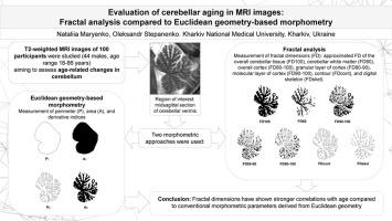
Two-dimensional T2-weighted magnetic resonance images from the brains of 100 conditionally healthy individuals (44 males and 56 females) aged 18–86 years were examined, with a focus on mid-sagittal sections of the cerebellar vermis. We determined ten parameters derived from Euclidean geometry (perimeter, area, and indices calculated from them), along with seven fractal dimension values derived from fractal geometry (the approximated fractal dimensions of the overall cerebellar tissue, white matter, overall cerebellar cortex and its granular and molecular layers, outer contour, and digital skeleton).
Results
Fractal dimensions demonstrated stronger correlation relationships with age compared to morphometric parameters derived from Euclidean geometry. The most pronounced age-related declines were observed in the approximated fractal dimensions of the cerebellar cortex and its layers, with decreases also noted in the fractal dimensions of the outer contour and digital cerebellar skeleton. Fractal dimension values did not significantly differ between males and females, while several Euclidean geometry-derived parameters showed sexual dimorphism. Although males demonstrated stronger relationships of some studied parameters with age, there was no statistically significant difference in the sex-related dynamics of aging.
Conclusion
The normal aging of the cerebellum involves not only absolute size alterations but also changes in the texture and spatial configuration of cerebellar tissue components, which can be quantitatively and objectively assessed by fractal analysis.

 求助内容:
求助内容: 应助结果提醒方式:


