Riccardo Bixio, Sara Bindoli, Andrea Morciano, Roberto Padoan, Federico Aldegheri, Francesca Mastropaolo, Eugenia Bertoldo, Denise Rotta, Matteo Appoloni, Giovanni Orsolini, Davide Gatti, Giovanni Adami, Ombretta Viapiana, Maurizio Rossini, Paolo Sfriso, Angelo Fassio
{"title":"The role of 18FDG–PET imaging in VEXAS syndrome: a multicentric case series and a systematic review of the literature","authors":"Riccardo Bixio, Sara Bindoli, Andrea Morciano, Roberto Padoan, Federico Aldegheri, Francesca Mastropaolo, Eugenia Bertoldo, Denise Rotta, Matteo Appoloni, Giovanni Orsolini, Davide Gatti, Giovanni Adami, Ombretta Viapiana, Maurizio Rossini, Paolo Sfriso, Angelo Fassio","doi":"10.1007/s11739-024-03763-9","DOIUrl":null,"url":null,"abstract":"<p>VEXAS (vacuoles, E1 enzyme, X-linked, autoinflammatory, and somatic) syndrome is characterized by heterogeneous clinical manifestations. Due to the inflammatory nature of this condition, 18-FDG–PET (18-fluorodeoxyglucose–positron emission tomography) might be used to diagnose and monitor the disease. However, no data are available about the most common findings of PET imaging in this disease. For this reason, we summarised all the available reports of patients with VEXAS who underwent at least one PET scan and described 8 additional patients’ PET from our centres. Overall, we described 35 patients’ PET findings. All patients were male, with a median age of 70 years. The most frequent hypermetabolic sites on PET scans were the bone marrow (77.1%), lymph nodes (35.3%), lungs (28.6%), spleen and large vessels (22.9%), and cartilage (20%). Six patients underwent a PET scan 2.7 ± 1.5 years before VEXAS diagnosis, showing nonspecific uptake in the bone marrow. Four patients had a follow-up PET scan, showing a decrease or a disappearance of the previously identified hypermetabolic areas. In conclusion, although no specific uptake site has been found for VEXAS syndrome, PET imaging could help detect inflammatory foci that are not clinically evident. In addition, high metabolic activity in bone marrow might precede the clinical onset of the disease, shedding light on the pathogenesis of VEXAS.</p>","PeriodicalId":13662,"journal":{"name":"Internal and Emergency Medicine","volume":null,"pages":null},"PeriodicalIF":3.2000,"publicationDate":"2024-09-09","publicationTypes":"Journal Article","fieldsOfStudy":null,"isOpenAccess":false,"openAccessPdf":"","citationCount":"0","resultStr":null,"platform":"Semanticscholar","paperid":null,"PeriodicalName":"Internal and Emergency Medicine","FirstCategoryId":"3","ListUrlMain":"https://doi.org/10.1007/s11739-024-03763-9","RegionNum":3,"RegionCategory":"医学","ArticlePicture":[],"TitleCN":null,"AbstractTextCN":null,"PMCID":null,"EPubDate":"","PubModel":"","JCR":"Q1","JCRName":"MEDICINE, GENERAL & INTERNAL","Score":null,"Total":0}
引用次数: 0
Abstract
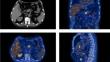
VEXAS (vacuoles, E1 enzyme, X-linked, autoinflammatory, and somatic) syndrome is characterized by heterogeneous clinical manifestations. Due to the inflammatory nature of this condition, 18-FDG–PET (18-fluorodeoxyglucose–positron emission tomography) might be used to diagnose and monitor the disease. However, no data are available about the most common findings of PET imaging in this disease. For this reason, we summarised all the available reports of patients with VEXAS who underwent at least one PET scan and described 8 additional patients’ PET from our centres. Overall, we described 35 patients’ PET findings. All patients were male, with a median age of 70 years. The most frequent hypermetabolic sites on PET scans were the bone marrow (77.1%), lymph nodes (35.3%), lungs (28.6%), spleen and large vessels (22.9%), and cartilage (20%). Six patients underwent a PET scan 2.7 ± 1.5 years before VEXAS diagnosis, showing nonspecific uptake in the bone marrow. Four patients had a follow-up PET scan, showing a decrease or a disappearance of the previously identified hypermetabolic areas. In conclusion, although no specific uptake site has been found for VEXAS syndrome, PET imaging could help detect inflammatory foci that are not clinically evident. In addition, high metabolic activity in bone marrow might precede the clinical onset of the disease, shedding light on the pathogenesis of VEXAS.
期刊介绍:
Internal and Emergency Medicine (IEM) is an independent, international, English-language, peer-reviewed journal designed for internists and emergency physicians. IEM publishes a variety of manuscript types including Original investigations, Review articles, Letters to the Editor, Editorials and Commentaries. Occasionally IEM accepts unsolicited Reviews, Commentaries or Editorials. The journal is divided into three sections, i.e., Internal Medicine, Emergency Medicine and Clinical Evidence and Health Technology Assessment, with three separate editorial boards. In the Internal Medicine section, invited Case records and Physical examinations, devoted to underlining the role of a clinical approach in selected clinical cases, are also published. The Emergency Medicine section will include a Morbidity and Mortality Report and an Airway Forum concerning the management of difficult airway problems. As far as Critical Care is becoming an integral part of Emergency Medicine, a new sub-section will report the literature that concerns the interface not only for the care of the critical patient in the Emergency Department, but also in the Intensive Care Unit. Finally, in the Clinical Evidence and Health Technology Assessment section brief discussions of topics of evidence-based medicine (Cochrane’s corner) and Research updates are published. IEM encourages letters of rebuttal and criticism of published articles. Topics of interest include all subjects that relate to the science and practice of Internal and Emergency Medicine.
 求助内容:
求助内容: 应助结果提醒方式:


