Nikolai V. Naoumov, David E. Kleiner, Elaine Chng, Dominique Brees, Chandra Saravanan, Yayun Ren, Dean Tai, Arun J. Sanyal
{"title":"Digital quantitation of bridging fibrosis and septa reveals changes in natural history and treatment not seen with conventional histology","authors":"Nikolai V. Naoumov, David E. Kleiner, Elaine Chng, Dominique Brees, Chandra Saravanan, Yayun Ren, Dean Tai, Arun J. Sanyal","doi":"10.1111/liv.16092","DOIUrl":null,"url":null,"abstract":"<div>\n \n \n <section>\n \n <h3> Background and Aims</h3>\n \n <p>Metabolic dysfunction-associated steatohepatitis (MASH) with bridging fibrosis is a critical stage in the evolution of fatty liver disease. Second harmonic generation/two-photon excitation fluorescence (SHG/TPEF) microscopy with artificial intelligence (AI) provides sensitive and reproducible quantitation of liver fibrosis. This methodology was applied to gain an in-depth understanding of intra-stage fibrosis changes and septa analyses in a homogenous, well-characterised group with MASH F3 fibrosis.</p>\n </section>\n \n <section>\n \n <h3> Methods</h3>\n \n <p>Paired liver biopsies (baseline [BL] and end of treatment [EOT]) of 57 patients (placebo, <i>n</i> = 17 and tropifexor <i>n</i> = 40), with F3 fibrosis stage at BL according to the clinical research network (CRN) scoring, were included. Unstained sections were examined using SHG/TPEF microscopy with AI. Changes in liver fibrosis overall and in five areas of liver lobules were quantitatively assessed by qFibrosis. Progressive, regressive septa, and 12 septa parameters were quantitatively analysed.</p>\n </section>\n \n <section>\n \n <h3> Results</h3>\n \n <p>qFibrosis demonstrated fibrosis progression or regression in 14/17 (82%) patients receiving placebo, while the CRN scoring categorised 11/17 (65%) as ‘no change’. Radar maps with qFibrosis readouts visualised quantitative fibrosis dynamics in different areas of liver lobules even in cases categorised as ‘No Change’. Measurement of septa parameters objectively differentiated regressive and progressive septa (<i>p</i> < .001). Quantitative changes in individual septa parameters (BL to EOT) were observed both in the ‘no change’ and the ‘regression’ subgroups, as defined by the CRN scoring.</p>\n </section>\n \n <section>\n \n <h3> Conclusion</h3>\n \n <p>SHG/TPEF microscopy with AI provides greater granularity and precision in assessing fibrosis dynamics in patients with bridging fibrosis, thus advancing knowledge development of fibrosis evolution in natural history and in clinical trials.</p>\n </section>\n </div>","PeriodicalId":18101,"journal":{"name":"Liver International","volume":"44 12","pages":"3214-3228"},"PeriodicalIF":6.0000,"publicationDate":"2024-09-09","publicationTypes":"Journal Article","fieldsOfStudy":null,"isOpenAccess":false,"openAccessPdf":"https://onlinelibrary.wiley.com/doi/epdf/10.1111/liv.16092","citationCount":"0","resultStr":null,"platform":"Semanticscholar","paperid":null,"PeriodicalName":"Liver International","FirstCategoryId":"3","ListUrlMain":"https://onlinelibrary.wiley.com/doi/10.1111/liv.16092","RegionNum":2,"RegionCategory":"医学","ArticlePicture":[],"TitleCN":null,"AbstractTextCN":null,"PMCID":null,"EPubDate":"","PubModel":"","JCR":"Q1","JCRName":"GASTROENTEROLOGY & HEPATOLOGY","Score":null,"Total":0}
引用次数: 0
Abstract
Background and Aims
Metabolic dysfunction-associated steatohepatitis (MASH) with bridging fibrosis is a critical stage in the evolution of fatty liver disease. Second harmonic generation/two-photon excitation fluorescence (SHG/TPEF) microscopy with artificial intelligence (AI) provides sensitive and reproducible quantitation of liver fibrosis. This methodology was applied to gain an in-depth understanding of intra-stage fibrosis changes and septa analyses in a homogenous, well-characterised group with MASH F3 fibrosis.
Methods
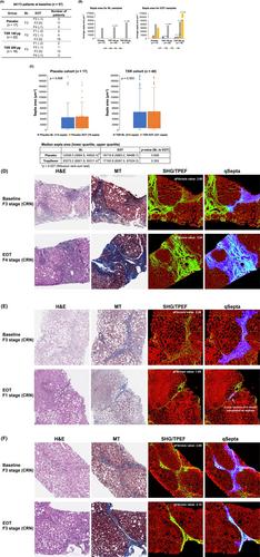
Paired liver biopsies (baseline [BL] and end of treatment [EOT]) of 57 patients (placebo, n = 17 and tropifexor n = 40), with F3 fibrosis stage at BL according to the clinical research network (CRN) scoring, were included. Unstained sections were examined using SHG/TPEF microscopy with AI. Changes in liver fibrosis overall and in five areas of liver lobules were quantitatively assessed by qFibrosis. Progressive, regressive septa, and 12 septa parameters were quantitatively analysed.
Results
qFibrosis demonstrated fibrosis progression or regression in 14/17 (82%) patients receiving placebo, while the CRN scoring categorised 11/17 (65%) as ‘no change’. Radar maps with qFibrosis readouts visualised quantitative fibrosis dynamics in different areas of liver lobules even in cases categorised as ‘No Change’. Measurement of septa parameters objectively differentiated regressive and progressive septa (p < .001). Quantitative changes in individual septa parameters (BL to EOT) were observed both in the ‘no change’ and the ‘regression’ subgroups, as defined by the CRN scoring.
Conclusion
SHG/TPEF microscopy with AI provides greater granularity and precision in assessing fibrosis dynamics in patients with bridging fibrosis, thus advancing knowledge development of fibrosis evolution in natural history and in clinical trials.
期刊介绍:
Liver International promotes all aspects of the science of hepatology from basic research to applied clinical studies. Providing an international forum for the publication of high-quality original research in hepatology, it is an essential resource for everyone working on normal and abnormal structure and function in the liver and its constituent cells, including clinicians and basic scientists involved in the multi-disciplinary field of hepatology. The journal welcomes articles from all fields of hepatology, which may be published as original articles, brief definitive reports, reviews, mini-reviews, images in hepatology and letters to the Editor.