Marc McLeod, Mario C Chang, Anna Rushin, Mukundan Ragavan, Rohit Mahar, Gaurav Sharma, Arshee Badar, Anthony Giacalone, Max E Glanz, Vinay R Malut, Dalton Graham, Nishanth E Sunny, James A Bankson, Kenneth Cusi, Matthew E Merritt
{"title":"Detecting altered hepatic lipid oxidation by MRI in an animal model of MASLD.","authors":"Marc McLeod, Mario C Chang, Anna Rushin, Mukundan Ragavan, Rohit Mahar, Gaurav Sharma, Arshee Badar, Anthony Giacalone, Max E Glanz, Vinay R Malut, Dalton Graham, Nishanth E Sunny, James A Bankson, Kenneth Cusi, Matthew E Merritt","doi":"10.1016/j.xcrm.2024.101714","DOIUrl":null,"url":null,"abstract":"<p><p>Metabolic dysfunction-associated steatotic liver disease (MASLD) prevalence is increasing annually and affects over a third of US adults. MASLD can progress to metabolic dysfunction-associated steatohepatitis (MASH), characterized by severe hepatocyte injury, inflammation, and eventual advanced fibrosis or cirrhosis. MASH is predicted to become the primary cause of liver transplant by 2030. Although the etiology of MASLD/MASH is incompletely understood, dysregulated fatty acid oxidation is implicated in disease pathogenesis. Here, we develop a method for estimating hepatic β-oxidation from the metabolism of [D<sub>15</sub>]octanoate to deuterated water and detection with deuterium magnetic resonance methods. Perfused livers from a mouse model of MASLD reveal dysregulated hepatic β-oxidation, findings that corroborate in vivo imaging. The high-fat-diet-induced MASLD mouse studies indicate that decreased β-oxidative efficiency in the fatty liver could serve as an indicator of MASLD progression. Furthermore, our method provides a clinically translatable imaging approach for determining hepatic β-oxidation efficiency.</p>","PeriodicalId":9822,"journal":{"name":"Cell Reports Medicine","volume":" ","pages":"101714"},"PeriodicalIF":11.7000,"publicationDate":"2024-09-17","publicationTypes":"Journal Article","fieldsOfStudy":null,"isOpenAccess":false,"openAccessPdf":"https://www.ncbi.nlm.nih.gov/pmc/articles/PMC11525016/pdf/","citationCount":"0","resultStr":null,"platform":"Semanticscholar","paperid":null,"PeriodicalName":"Cell Reports Medicine","FirstCategoryId":"3","ListUrlMain":"https://doi.org/10.1016/j.xcrm.2024.101714","RegionNum":1,"RegionCategory":"医学","ArticlePicture":[],"TitleCN":null,"AbstractTextCN":null,"PMCID":null,"EPubDate":"2024/9/5 0:00:00","PubModel":"Epub","JCR":"Q1","JCRName":"CELL BIOLOGY","Score":null,"Total":0}
引用次数: 0
Abstract
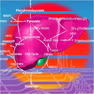
Metabolic dysfunction-associated steatotic liver disease (MASLD) prevalence is increasing annually and affects over a third of US adults. MASLD can progress to metabolic dysfunction-associated steatohepatitis (MASH), characterized by severe hepatocyte injury, inflammation, and eventual advanced fibrosis or cirrhosis. MASH is predicted to become the primary cause of liver transplant by 2030. Although the etiology of MASLD/MASH is incompletely understood, dysregulated fatty acid oxidation is implicated in disease pathogenesis. Here, we develop a method for estimating hepatic β-oxidation from the metabolism of [D15]octanoate to deuterated water and detection with deuterium magnetic resonance methods. Perfused livers from a mouse model of MASLD reveal dysregulated hepatic β-oxidation, findings that corroborate in vivo imaging. The high-fat-diet-induced MASLD mouse studies indicate that decreased β-oxidative efficiency in the fatty liver could serve as an indicator of MASLD progression. Furthermore, our method provides a clinically translatable imaging approach for determining hepatic β-oxidation efficiency.
Cell Reports MedicineBiochemistry, Genetics and Molecular Biology-Biochemistry, Genetics and Molecular Biology (all)
CiteScore
15.00
自引率
1.40%
发文量
231
审稿时长
40 days
期刊介绍:
Cell Reports Medicine is an esteemed open-access journal by Cell Press that publishes groundbreaking research in translational and clinical biomedical sciences, influencing human health and medicine.
Our journal ensures wide visibility and accessibility, reaching scientists and clinicians across various medical disciplines. We publish original research that spans from intriguing human biology concepts to all aspects of clinical work. We encourage submissions that introduce innovative ideas, forging new paths in clinical research and practice. We also welcome studies that provide vital information, enhancing our understanding of current standards of care in diagnosis, treatment, and prognosis. This encompasses translational studies, clinical trials (including long-term follow-ups), genomics, biomarker discovery, and technological advancements that contribute to diagnostics, treatment, and healthcare. Additionally, studies based on vertebrate model organisms are within the scope of the journal, as long as they directly relate to human health and disease.
 求助内容:
求助内容: 应助结果提醒方式:


