Differences in Topography of Individual Amyloid Brain Networks by Amyloid PET Images in Healthy Control, Mild Cognitive Impairment, and Alzheimer's Disease.
{"title":"Differences in Topography of Individual Amyloid Brain Networks by Amyloid PET Images in Healthy Control, Mild Cognitive Impairment, and Alzheimer's Disease.","authors":"Tsung-Ying Ho, Shu-Hua Huang, Chi-Wei Huang, Kun-Ju Lin, Jung-Lung Hsu, Kuo-Lun Huang, Ko-Ting Chen, Chiung-Chih Chang, Ing-Tsung Hsiao, Sheng-Yao Huang","doi":"10.1007/s10278-024-01230-7","DOIUrl":null,"url":null,"abstract":"<p><p>Amyloid plaques, implicated in Alzheimer's disease, exhibit a spatial propagation pattern through interconnected brain regions, suggesting network-driven dissemination. This study utilizes PET imaging to investigate these brain connections and introduces an innovative method for analyzing the amyloid network. A modified version of a previously established method is applied to explore distinctive patterns of connectivity alterations across cognitive performance domains. PET images illustrate differences in amyloid accumulation, complemented by quantitative network indices. The normal control group shows minimal amyloid accumulation and preserved network connectivity. The MCI group displays intermediate amyloid deposits and partial similarity to normal controls and AD patients, reflecting the evolving nature of cognitive decline. Alzheimer's disease patients exhibit high amyloid levels and pronounced disruptions in network connectivity, which are reflected in low levels of global efficiency (Eg) and local efficiency (Eloc). It is mostly in the temporal lobe where connectivity alterations are found, particularly in regions related to memory and cognition. Network connectivity alterations, combined with amyloid PET imaging, show potential as discriminative markers for different cognitive states. Dataset-specific variations must be considered when interpreting connectivity patterns. The variability in MCI and AD overlap emphasizes the heterogeneity in cognitive decline progression, suggesting personalized approaches for neurodegenerative disorders. This study contributes to understanding the evolving network characteristics associated with normal cognition, MCI, and AD, offering valuable insights for developing diagnostic and prognostic markers.</p>","PeriodicalId":516858,"journal":{"name":"Journal of imaging informatics in medicine","volume":" ","pages":"681-693"},"PeriodicalIF":0.0000,"publicationDate":"2025-04-01","publicationTypes":"Journal Article","fieldsOfStudy":null,"isOpenAccess":false,"openAccessPdf":"https://www.ncbi.nlm.nih.gov/pmc/articles/PMC11950497/pdf/","citationCount":"0","resultStr":null,"platform":"Semanticscholar","paperid":null,"PeriodicalName":"Journal of imaging informatics in medicine","FirstCategoryId":"1085","ListUrlMain":"https://doi.org/10.1007/s10278-024-01230-7","RegionNum":0,"RegionCategory":null,"ArticlePicture":[],"TitleCN":null,"AbstractTextCN":null,"PMCID":null,"EPubDate":"2024/9/4 0:00:00","PubModel":"Epub","JCR":"","JCRName":"","Score":null,"Total":0}
引用次数: 0
Abstract
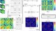
Amyloid plaques, implicated in Alzheimer's disease, exhibit a spatial propagation pattern through interconnected brain regions, suggesting network-driven dissemination. This study utilizes PET imaging to investigate these brain connections and introduces an innovative method for analyzing the amyloid network. A modified version of a previously established method is applied to explore distinctive patterns of connectivity alterations across cognitive performance domains. PET images illustrate differences in amyloid accumulation, complemented by quantitative network indices. The normal control group shows minimal amyloid accumulation and preserved network connectivity. The MCI group displays intermediate amyloid deposits and partial similarity to normal controls and AD patients, reflecting the evolving nature of cognitive decline. Alzheimer's disease patients exhibit high amyloid levels and pronounced disruptions in network connectivity, which are reflected in low levels of global efficiency (Eg) and local efficiency (Eloc). It is mostly in the temporal lobe where connectivity alterations are found, particularly in regions related to memory and cognition. Network connectivity alterations, combined with amyloid PET imaging, show potential as discriminative markers for different cognitive states. Dataset-specific variations must be considered when interpreting connectivity patterns. The variability in MCI and AD overlap emphasizes the heterogeneity in cognitive decline progression, suggesting personalized approaches for neurodegenerative disorders. This study contributes to understanding the evolving network characteristics associated with normal cognition, MCI, and AD, offering valuable insights for developing diagnostic and prognostic markers.
通过淀粉样蛋白 PET 图像观察健康对照组、轻度认知障碍患者和阿尔茨海默氏症患者个体淀粉样蛋白脑网络拓扑结构的差异。
与阿尔茨海默病有关的淀粉样蛋白斑块在相互连接的脑区中呈现出空间传播模式,这表明淀粉样蛋白斑块的传播是由网络驱动的。本研究利用正电子发射计算机断层成像技术研究这些大脑连接,并引入了一种创新的淀粉样蛋白网络分析方法。该方法是对以前建立的方法的改进版,用于探索不同认知能力领域的连接改变的独特模式。PET 图像显示了淀粉样蛋白积累的差异,并辅以定量网络指数。正常对照组显示出最小的淀粉样蛋白积累和保留的网络连通性。MCI 组显示出中等程度的淀粉样蛋白沉积,与正常对照组和阿兹海默症患者部分相似,反映出认知功能衰退的演变性质。阿尔茨海默病患者的淀粉样蛋白水平较高,网络连通性受到明显破坏,这反映在全局效率(Eg)和局部效率(Eloc)水平较低。连接性改变主要发生在颞叶,尤其是与记忆和认知相关的区域。网络连通性改变与淀粉样蛋白 PET 成像相结合,显示出作为不同认知状态判别标志物的潜力。在解释连通性模式时,必须考虑数据集的特定差异。MCI和AD重叠的变异性强调了认知功能衰退进展的异质性,建议采用个性化方法治疗神经退行性疾病。这项研究有助于了解与正常认知、MCI 和 AD 相关的不断变化的网络特征,为开发诊断和预后标记物提供了宝贵的见解。

 求助内容:
求助内容: 应助结果提醒方式:


