Neural network segmentation of disc volume from magnetic resonance images and the effect of degeneration and spinal level

IF 3.4 3区 医学 Q1 ORTHOPEDICS
JOR Spine Pub Date : 2024-09-04 DOI:10.1002/jsp2.70000
Milad I. Markhali, John M. Peloquin, Kyle D. Meadows, Harrah R. Newman, Dawn M. Elliott
{"title":"Neural network segmentation of disc volume from magnetic resonance images and the effect of degeneration and spinal level","authors":"Milad I. Markhali,&nbsp;John M. Peloquin,&nbsp;Kyle D. Meadows,&nbsp;Harrah R. Newman,&nbsp;Dawn M. Elliott","doi":"10.1002/jsp2.70000","DOIUrl":null,"url":null,"abstract":"<div>\n \n \n <section>\n \n <h3> Background</h3>\n \n <p>Magnetic resonance imaging (MRI) noninvasively quantifies disc structure but requires segmentation that is both time intensive and susceptible to human error. Recent advances in neural networks can improve on manual segmentation. The aim of this study was to establish a method for automatic slice-wise segmentation of 3D disc volumes from subjects with a wide range of age and degrees of disc degeneration. A U-Net convolutional neural network was trained to segment 3D T1-weighted spine MRI.</p>\n </section>\n \n <section>\n \n <h3> Methods</h3>\n \n <p>Lumbar spine MRIs were acquired from 43 subjects (23–83 years old) and manually segmented. A U-Net architecture was trained using the TensorFlow framework. Two rounds of model tuning were performed. The performance of the model was measured using a validation set that did not cross over from the training set. The model version with the best Dice similarity coefficient (DSC) was selected in each tuning round. After model development was complete and a final U-Net model was selected, performance of this model was compared between disc levels and degeneration grades.</p>\n </section>\n \n <section>\n \n <h3> Results</h3>\n \n <p>Performance of the final model was equivalent to manual segmentation, with a mean DSC = 0.935 ± 0.014 for degeneration grades I–IV. Neither the manual segmentation nor the U-Net model performed as well for grade V disc segmentation. Compared with the baseline model at the beginning of round 1, the best model had fewer filters/parameters (75%), was trained using only slices with at least one disc-labeled pixel, applied contrast stretching to its input images, and used a greater dropout rate.</p>\n </section>\n \n <section>\n \n <h3> Conclusion</h3>\n \n <p>This study successfully trained a U-Net model for automatic slice-wise segmentation of 3D disc volumes from populations with a wide range of ages and disc degeneration. The final trained model is available to support scientific use.</p>\n </section>\n </div>","PeriodicalId":14876,"journal":{"name":"JOR Spine","volume":null,"pages":null},"PeriodicalIF":3.4000,"publicationDate":"2024-09-04","publicationTypes":"Journal Article","fieldsOfStudy":null,"isOpenAccess":false,"openAccessPdf":"https://www.ncbi.nlm.nih.gov/pmc/articles/PMC11372286/pdf/","citationCount":"0","resultStr":null,"platform":"Semanticscholar","paperid":null,"PeriodicalName":"JOR Spine","FirstCategoryId":"3","ListUrlMain":"https://onlinelibrary.wiley.com/doi/10.1002/jsp2.70000","RegionNum":3,"RegionCategory":"医学","ArticlePicture":[],"TitleCN":null,"AbstractTextCN":null,"PMCID":null,"EPubDate":"","PubModel":"","JCR":"Q1","JCRName":"ORTHOPEDICS","Score":null,"Total":0}
引用次数: 0

Abstract

Background

Magnetic resonance imaging (MRI) noninvasively quantifies disc structure but requires segmentation that is both time intensive and susceptible to human error. Recent advances in neural networks can improve on manual segmentation. The aim of this study was to establish a method for automatic slice-wise segmentation of 3D disc volumes from subjects with a wide range of age and degrees of disc degeneration. A U-Net convolutional neural network was trained to segment 3D T1-weighted spine MRI.

Methods

Lumbar spine MRIs were acquired from 43 subjects (23–83 years old) and manually segmented. A U-Net architecture was trained using the TensorFlow framework. Two rounds of model tuning were performed. The performance of the model was measured using a validation set that did not cross over from the training set. The model version with the best Dice similarity coefficient (DSC) was selected in each tuning round. After model development was complete and a final U-Net model was selected, performance of this model was compared between disc levels and degeneration grades.

Results

Performance of the final model was equivalent to manual segmentation, with a mean DSC = 0.935 ± 0.014 for degeneration grades I–IV. Neither the manual segmentation nor the U-Net model performed as well for grade V disc segmentation. Compared with the baseline model at the beginning of round 1, the best model had fewer filters/parameters (75%), was trained using only slices with at least one disc-labeled pixel, applied contrast stretching to its input images, and used a greater dropout rate.

Conclusion

This study successfully trained a U-Net model for automatic slice-wise segmentation of 3D disc volumes from populations with a wide range of ages and disc degeneration. The final trained model is available to support scientific use.
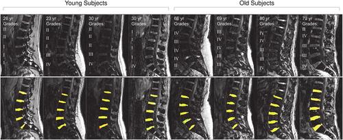
Abstract Image

神经网络分割磁共振图像中的椎间盘体积以及退化和脊柱水平的影响。
背景:磁共振成像(MRI)可无创量化椎间盘结构,但需要进行既耗时又容易出现人为错误的分割。神经网络的最新进展可以改进人工分割。本研究的目的是建立一种方法,对年龄和椎间盘退化程度不同的受试者的三维椎间盘体积进行自动切片分割。对 U-Net 卷积神经网络进行了训练,以分割三维 T1 加权脊柱 MRI:方法:采集了 43 名受试者(23-83 岁)的腰椎 MRI 图像,并进行手动分割。使用 TensorFlow 框架训练 U-Net 架构。对模型进行了两轮调整。模型的性能是通过一个不与训练集交叉的验证集来测量的。在每一轮调整中,都会选择具有最佳骰子相似系数(DSC)的模型版本。模型开发完成并选出最终的 U-Net 模型后,对该模型在不同椎间盘水平和退变等级之间的性能进行了比较:结果:最终模型的性能与人工分割相当,I-IV 级退变的平均 DSC = 0.935 ± 0.014。人工分割和 U-Net 模型在 V 级椎间盘分割中的表现都不理想。与第一轮开始时的基线模型相比,最佳模型的过滤器/参数较少(75%),仅使用至少有一个椎间盘标记像素的切片进行训练,对输入图像进行对比度拉伸,并且使用了更高的辍学率:这项研究成功地训练了一个 U-Net 模型,该模型可对年龄和椎间盘退变程度不同的人群的三维椎间盘体积进行自动切片分割。最终训练出的模型可用于科学研究。
本文章由计算机程序翻译,如有差异,请以英文原文为准。
求助全文
约1分钟内获得全文 求助全文
来源期刊
JOR Spine
JOR Spine ORTHOPEDICS-
CiteScore
6.40
自引率
18.90%
发文量
42
审稿时长
10 weeks
×
引用
GB/T 7714-2015
复制
MLA
复制
APA
复制
导出至
BibTeX EndNote RefMan NoteFirst NoteExpress
×
提示
您的信息不完整,为了账户安全,请先补充。
现在去补充
×
提示
您因"违规操作"
具体请查看互助需知
我知道了
×
提示
确定
请完成安全验证×
copy
已复制链接
快去分享给好友吧!
我知道了
右上角分享
点击右上角分享
0
联系我们:info@booksci.cn Book学术提供免费学术资源搜索服务,方便国内外学者检索中英文文献。致力于提供最便捷和优质的服务体验。 Copyright © 2023 布克学术 All rights reserved.
京ICP备2023020795号-1
ghs 京公网安备 11010802042870号
Book学术文献互助
Book学术文献互助群
群 号:481959085
Book学术官方微信