Pedram Mahmoudi Aliabadi, Khlowd Al-Qaisi, Vishnu Reddy, Andreas Radbruch, Makio Kobayashi, Hiromi Kubagawa
{"title":"Periodic Acid Schiff Staining to Detect Mott Cells, Aberrant Plasma Cells Containing Immunoglobulin Inclusions Called Russell Bodies","authors":"Pedram Mahmoudi Aliabadi, Khlowd Al-Qaisi, Vishnu Reddy, Andreas Radbruch, Makio Kobayashi, Hiromi Kubagawa","doi":"10.1002/cpz1.70005","DOIUrl":null,"url":null,"abstract":"<p>Hematoxylin and eosin staining is widely used for routine histopathological analysis under light microscopic examination to determine alterations of tissue architecture and cellular components in animal studies. Aside from hematoxylin/eosin staining, periodic acid Schiff (PAS) staining is used to detect polysaccharides and carbohydrate-rich macromolecules, and is essential in immunological fields for evaluation of glomerular lesions of kidneys in autoimmune animals. Since erythrocytes are not stained by PAS, this stain is also helpful for identifying changes in immune cells in the red pulp of the spleen, which is filled with erythrocytes. This article describes a protocol to detect Mott cells, bizarre plasma cells containing immunoglobulin inclusion bodies (Russell bodies) in the cytoplasm. The protocol can be used for formalin-fixed, paraffin-embedded tissue sections, frozen tissue sections, tissue-touch preparations, blood films, and cytocentrifuged cell smears. © 2024 The Author(s). Current Protocols published by Wiley Periodicals LLC.</p><p><b>Basic Protocol 1</b>: Detection of Mott cells by PAS staining in formalin-fixed, paraffin-embedded tissue sections</p><p><b>Basic Protocol 2</b>: Detection of Mott cells by PAS staining in frozen tissue sections, touch preparations, blood films, and cytocentrifuged cell smears</p>","PeriodicalId":93970,"journal":{"name":"Current protocols","volume":"4 9","pages":""},"PeriodicalIF":0.0000,"publicationDate":"2024-09-04","publicationTypes":"Journal Article","fieldsOfStudy":null,"isOpenAccess":false,"openAccessPdf":"https://onlinelibrary.wiley.com/doi/epdf/10.1002/cpz1.70005","citationCount":"0","resultStr":null,"platform":"Semanticscholar","paperid":null,"PeriodicalName":"Current protocols","FirstCategoryId":"1085","ListUrlMain":"https://onlinelibrary.wiley.com/doi/10.1002/cpz1.70005","RegionNum":0,"RegionCategory":null,"ArticlePicture":[],"TitleCN":null,"AbstractTextCN":null,"PMCID":null,"EPubDate":"","PubModel":"","JCR":"","JCRName":"","Score":null,"Total":0}
引用次数: 0
Abstract
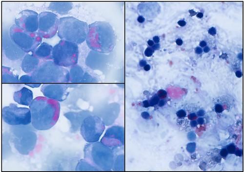
Hematoxylin and eosin staining is widely used for routine histopathological analysis under light microscopic examination to determine alterations of tissue architecture and cellular components in animal studies. Aside from hematoxylin/eosin staining, periodic acid Schiff (PAS) staining is used to detect polysaccharides and carbohydrate-rich macromolecules, and is essential in immunological fields for evaluation of glomerular lesions of kidneys in autoimmune animals. Since erythrocytes are not stained by PAS, this stain is also helpful for identifying changes in immune cells in the red pulp of the spleen, which is filled with erythrocytes. This article describes a protocol to detect Mott cells, bizarre plasma cells containing immunoglobulin inclusion bodies (Russell bodies) in the cytoplasm. The protocol can be used for formalin-fixed, paraffin-embedded tissue sections, frozen tissue sections, tissue-touch preparations, blood films, and cytocentrifuged cell smears. © 2024 The Author(s). Current Protocols published by Wiley Periodicals LLC.
Basic Protocol 1: Detection of Mott cells by PAS staining in formalin-fixed, paraffin-embedded tissue sections
Basic Protocol 2: Detection of Mott cells by PAS staining in frozen tissue sections, touch preparations, blood films, and cytocentrifuged cell smears

周期性酸性希夫染色法检测莫特细胞(Mott Cells),一种含有被称为罗素体的免疫球蛋白包涵体的异常浆细胞。
在动物研究中,苏木精和伊红染色被广泛用于光镜下的常规组织病理学分析,以确定组织结构和细胞成分的改变。除苏木精/伊红染色外,周期性酸性希夫(PAS)染色也用于检测多糖和富含碳水化合物的大分子,是免疫学领域评估自身免疫性动物肾小球病变的重要方法。由于红细胞不能被 PAS 染色,因此这种染色法也有助于鉴别充满红细胞的脾脏红髓中免疫细胞的变化。本文介绍了一种检测莫特细胞(细胞质中含有免疫球蛋白包涵体(罗素体)的奇异浆细胞)的方案。该方案可用于福尔马林固定、石蜡包埋的组织切片、冷冻组织切片、组织触片制备、血片和细胞离心细胞涂片。© 2024 作者。当前协议》由 Wiley Periodicals LLC 出版。基本方案 1:通过对福尔马林固定、石蜡包埋的组织切片进行 PAS 染色来检测莫特细胞 基本方案 2:通过对冷冻组织切片、接触制备物、血片和细胞离心涂片进行 PAS 染色来检测莫特细胞。
本文章由计算机程序翻译,如有差异,请以英文原文为准。


 求助内容:
求助内容: 应助结果提醒方式:


