Median nerve lesions in pediatric displaced supracondylar humerus fracture: A prospective neurological, electrodiagnostic and ultrasound characterization
Ines Sophie Schädlich, Sabriena Buschbaum, Tim Magnus, Konrad Reinshagen, Kristofer Wintges, Mathias Gelderblom
{"title":"Median nerve lesions in pediatric displaced supracondylar humerus fracture: A prospective neurological, electrodiagnostic and ultrasound characterization","authors":"Ines Sophie Schädlich, Sabriena Buschbaum, Tim Magnus, Konrad Reinshagen, Kristofer Wintges, Mathias Gelderblom","doi":"10.1111/ene.16459","DOIUrl":null,"url":null,"abstract":"<div>\n \n \n <section>\n \n <h3> Background and Purpose</h3>\n \n <p>Supracondylar humerus fractures (SCHFs) are the most common elbow fractures in children. Traumatic median nerve injury and isolated lesions of its pure forearm motor branch, anterior interosseus nerve (AIN), have both been independently reported as complications of displaced SCHFs. Our main objectives were to characterize the neurological syndrome to distinguish median nerve from AIN lesions and to determine the prognosis of median nerve lesions after displaced SCHFs.</p>\n </section>\n \n <section>\n \n <h3> Methods</h3>\n \n <p>Ten children were prospectively followed for an average of 11.6 months. Patients received a standardized clinical examination and high-resolution ultrasound of the median nerve every 1–3 months starting 1–2 months after trauma. Electrodiagnostic studies were performed within the first 4 months and after complete clinical recovery.</p>\n </section>\n \n <section>\n \n <h3> Results</h3>\n \n <p>All children shared a clinical syndrome with predominant but not exclusive affection of AIN innervated muscles. High-resolution ultrasound uniformly excluded persistent nerve entrapment and neurotmesis requiring revision surgery but visualized post-traumatic median nerve neuroma at the fracture site in all patients. Electrodiagnostic studies showed axonal motor and sensory median nerve neuropathy. All children achieved complete functional recovery under conservative management. Motor recovery required up to 11 months and differed between involved muscles.</p>\n </section>\n \n <section>\n \n <h3> Conclusions</h3>\n \n <p>It was shown that neurological deficits of the median nerve in displaced SCHFs exceeded an isolated AIN lesion. Notably, detailed neurological follow-up examinations and sonographic exclusion of persistent nerve compression were able to guide conservative therapy in affected children. Under these conditions the prognosis of median nerve lesions was excellent despite severe initial deficits, development of neuroma and axonal injury.</p>\n </section>\n </div>","PeriodicalId":11954,"journal":{"name":"European Journal of Neurology","volume":"31 12","pages":""},"PeriodicalIF":4.5000,"publicationDate":"2024-09-04","publicationTypes":"Journal Article","fieldsOfStudy":null,"isOpenAccess":false,"openAccessPdf":"https://www.ncbi.nlm.nih.gov/pmc/articles/PMC11555132/pdf/","citationCount":"0","resultStr":null,"platform":"Semanticscholar","paperid":null,"PeriodicalName":"European Journal of Neurology","FirstCategoryId":"3","ListUrlMain":"https://onlinelibrary.wiley.com/doi/10.1111/ene.16459","RegionNum":2,"RegionCategory":"医学","ArticlePicture":[],"TitleCN":null,"AbstractTextCN":null,"PMCID":null,"EPubDate":"","PubModel":"","JCR":"Q1","JCRName":"CLINICAL NEUROLOGY","Score":null,"Total":0}
引用次数: 0
Abstract
Background and Purpose
Supracondylar humerus fractures (SCHFs) are the most common elbow fractures in children. Traumatic median nerve injury and isolated lesions of its pure forearm motor branch, anterior interosseus nerve (AIN), have both been independently reported as complications of displaced SCHFs. Our main objectives were to characterize the neurological syndrome to distinguish median nerve from AIN lesions and to determine the prognosis of median nerve lesions after displaced SCHFs.
Methods
Ten children were prospectively followed for an average of 11.6 months. Patients received a standardized clinical examination and high-resolution ultrasound of the median nerve every 1–3 months starting 1–2 months after trauma. Electrodiagnostic studies were performed within the first 4 months and after complete clinical recovery.
Results
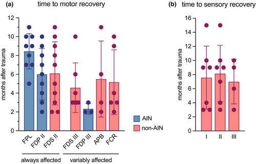
All children shared a clinical syndrome with predominant but not exclusive affection of AIN innervated muscles. High-resolution ultrasound uniformly excluded persistent nerve entrapment and neurotmesis requiring revision surgery but visualized post-traumatic median nerve neuroma at the fracture site in all patients. Electrodiagnostic studies showed axonal motor and sensory median nerve neuropathy. All children achieved complete functional recovery under conservative management. Motor recovery required up to 11 months and differed between involved muscles.
Conclusions
It was shown that neurological deficits of the median nerve in displaced SCHFs exceeded an isolated AIN lesion. Notably, detailed neurological follow-up examinations and sonographic exclusion of persistent nerve compression were able to guide conservative therapy in affected children. Under these conditions the prognosis of median nerve lesions was excellent despite severe initial deficits, development of neuroma and axonal injury.
期刊介绍:
The European Journal of Neurology is the official journal of the European Academy of Neurology and covers all areas of clinical and basic research in neurology, including pre-clinical research of immediate translational value for new potential treatments. Emphasis is placed on major diseases of large clinical and socio-economic importance (dementia, stroke, epilepsy, headache, multiple sclerosis, movement disorders, and infectious diseases).