{"title":"Evaluation of micro-remnant niduses of arteriovenous malformations post-gamma knife radiosurgery by 3D-rotational angiography","authors":"Ryuichi Noda, Atsuya Akabane, Mariko Kawashima, Masafumi Segawa, Sho Tsunoda, Hiroyuki Wada, Makoto Watanabe, Haruyasu Yamada, Tomohiro Inoue","doi":"10.1007/s00701-024-06246-0","DOIUrl":null,"url":null,"abstract":"<div><h3>Purpose</h3><p>Recent innovations in radiological imaging have enabled the detection of micro-remnant niduses of arteriovenous malformations (AVMs) after gamma knife radiosurgery (GKS), which have not been previously perceptible. Herein, we focus on the difficulty of evaluating micro-remnant AVMs after GKS that are hardly perceptible on conventional examinations and propose integrating follow-up three-dimensional rotational angiography (3D-RA) in the previous gamma plan as a solution.</p><h3>Methods</h3><p>We retrospectively searched NTT Medical Center Tokyo hospital database for patients with AVMs who underwent both two-dimensional digital subtraction angiography (2D-DSA) and 3D-RA as follow-up for GKS from February 2021 to January 2024. Patients with suspected nidus occlusion on the latest non-contrast-enhanced magnetic resonance angiography (NC-MRA) were included, and contrast-enhanced magnetic resonance angiography (CE-MRA), 2D-DSA, and 3D-RA were evaluated.</p><h3>Results</h3><p>Twelve patients with 13 AVM sites were defined as having complete nidus occlusion on upfront NC-MRA. On 2D-DSA, seven AVM sites showed the presence of slight remaining AVMs based on the detection of remnant drainage veins, however the nidus was not detected in three cases. Nevertheless, 3D-RA detected micro-remnant niduses in all seven AVM sites, and four patients underwent re-GKS. Nine patients with ten AVM sites also underwent CE-MRA, and six AVM sites were diagnosed with radiation-induced parenchymal injury.</p><h3>Conclusion</h3><p>Importing the 3D-RA image into the treatment planning has the potential to be more helpful than NC-MRA or CE-MRA to detect micro-remnant AVMs and evaluate the true remnant volume, and may contribute to a more detailed treatment planning, thereby improving the results of GKS retreatment.</p></div>","PeriodicalId":7370,"journal":{"name":"Acta Neurochirurgica","volume":"166 1","pages":""},"PeriodicalIF":1.9000,"publicationDate":"2024-09-03","publicationTypes":"Journal Article","fieldsOfStudy":null,"isOpenAccess":false,"openAccessPdf":"","citationCount":"0","resultStr":null,"platform":"Semanticscholar","paperid":null,"PeriodicalName":"Acta Neurochirurgica","FirstCategoryId":"3","ListUrlMain":"https://link.springer.com/article/10.1007/s00701-024-06246-0","RegionNum":3,"RegionCategory":"医学","ArticlePicture":[],"TitleCN":null,"AbstractTextCN":null,"PMCID":null,"EPubDate":"","PubModel":"","JCR":"Q3","JCRName":"CLINICAL NEUROLOGY","Score":null,"Total":0}
引用次数: 0
Abstract
Purpose
Recent innovations in radiological imaging have enabled the detection of micro-remnant niduses of arteriovenous malformations (AVMs) after gamma knife radiosurgery (GKS), which have not been previously perceptible. Herein, we focus on the difficulty of evaluating micro-remnant AVMs after GKS that are hardly perceptible on conventional examinations and propose integrating follow-up three-dimensional rotational angiography (3D-RA) in the previous gamma plan as a solution.
Methods
We retrospectively searched NTT Medical Center Tokyo hospital database for patients with AVMs who underwent both two-dimensional digital subtraction angiography (2D-DSA) and 3D-RA as follow-up for GKS from February 2021 to January 2024. Patients with suspected nidus occlusion on the latest non-contrast-enhanced magnetic resonance angiography (NC-MRA) were included, and contrast-enhanced magnetic resonance angiography (CE-MRA), 2D-DSA, and 3D-RA were evaluated.
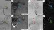
Results
Twelve patients with 13 AVM sites were defined as having complete nidus occlusion on upfront NC-MRA. On 2D-DSA, seven AVM sites showed the presence of slight remaining AVMs based on the detection of remnant drainage veins, however the nidus was not detected in three cases. Nevertheless, 3D-RA detected micro-remnant niduses in all seven AVM sites, and four patients underwent re-GKS. Nine patients with ten AVM sites also underwent CE-MRA, and six AVM sites were diagnosed with radiation-induced parenchymal injury.
Conclusion
Importing the 3D-RA image into the treatment planning has the potential to be more helpful than NC-MRA or CE-MRA to detect micro-remnant AVMs and evaluate the true remnant volume, and may contribute to a more detailed treatment planning, thereby improving the results of GKS retreatment.
期刊介绍:
The journal "Acta Neurochirurgica" publishes only original papers useful both to research and clinical work. Papers should deal with clinical neurosurgery - diagnosis and diagnostic techniques, operative surgery and results, postoperative treatment - or with research work in neuroscience if the underlying questions or the results are of neurosurgical interest. Reports on congresses are given in brief accounts. As official organ of the European Association of Neurosurgical Societies the journal publishes all announcements of the E.A.N.S. and reports on the activities of its member societies. Only contributions written in English will be accepted.

 求助内容:
求助内容: 应助结果提醒方式:


