Fabio Galbusera, Andrea Cina, Dave O'Riordan, Jacopo A Vitale, Markus Loibl, Tamás F Fekete, Frank Kleinstück, Daniel Haschtmann, Anne F Mannion
{"title":"Estimating lumbar bone mineral density from conventional MRI and radiographs with deep learning in spine patients.","authors":"Fabio Galbusera, Andrea Cina, Dave O'Riordan, Jacopo A Vitale, Markus Loibl, Tamás F Fekete, Frank Kleinstück, Daniel Haschtmann, Anne F Mannion","doi":"10.1007/s00586-024-08463-8","DOIUrl":null,"url":null,"abstract":"<p><strong>Purpose: </strong>This study aimed to develop machine learning methods to estimate bone mineral density and detect osteopenia/osteoporosis from conventional lumbar MRI (T1-weighted and T2-weighted images) and planar radiography in combination with clinical data and imaging parameters of the acquisition protocol.</p><p><strong>Methods: </strong>A database of 429 patients subjected to lumbar MRI, radiographs and dual-energy x-ray absorptiometry within 6 months was created from an institutional database. Several machine learning models were trained and tested (373 patients for training, 86 for testing) with the following objectives: (1) direct estimation of the vertebral bone mineral density; (2) classification of T-score lower than - 1 or (3) lower than - 2.5. The models took as inputs either the images or radiomics features derived from them, alone or in combination with metadata (age, sex, body size, vertebral level, parameters of the imaging protocol).</p><p><strong>Results: </strong>The best-performing models achieved mean absolute errors of 0.15-0.16 g/cm<sup>2</sup> for the direct estimation of bone mineral density, and areas under the receiver operating characteristic curve of 0.82 (MRIs) - 0.80 (radiographs) for the classification of T-scores lower than - 1, and 0.80 (MRIs) - 0.65 (radiographs) for T-scores lower than - 2.5.</p><p><strong>Conclusions: </strong>The models showed good discriminative performances in detecting cases of low bone mineral density, and more limited capabilities for the direct estimation of its value. Being based on routine imaging and readily available data, such models are promising tools to retrospectively analyse existing datasets as well as for the opportunistic investigation of bone disorders.</p>","PeriodicalId":12323,"journal":{"name":"European Spine Journal","volume":" ","pages":"4092-4103"},"PeriodicalIF":2.6000,"publicationDate":"2024-11-01","publicationTypes":"Journal Article","fieldsOfStudy":null,"isOpenAccess":false,"openAccessPdf":"","citationCount":"0","resultStr":null,"platform":"Semanticscholar","paperid":null,"PeriodicalName":"European Spine Journal","FirstCategoryId":"3","ListUrlMain":"https://doi.org/10.1007/s00586-024-08463-8","RegionNum":3,"RegionCategory":"医学","ArticlePicture":[],"TitleCN":null,"AbstractTextCN":null,"PMCID":null,"EPubDate":"2024/8/30 0:00:00","PubModel":"Epub","JCR":"Q2","JCRName":"CLINICAL NEUROLOGY","Score":null,"Total":0}
引用次数: 0
Abstract
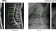
Purpose: This study aimed to develop machine learning methods to estimate bone mineral density and detect osteopenia/osteoporosis from conventional lumbar MRI (T1-weighted and T2-weighted images) and planar radiography in combination with clinical data and imaging parameters of the acquisition protocol.
Methods: A database of 429 patients subjected to lumbar MRI, radiographs and dual-energy x-ray absorptiometry within 6 months was created from an institutional database. Several machine learning models were trained and tested (373 patients for training, 86 for testing) with the following objectives: (1) direct estimation of the vertebral bone mineral density; (2) classification of T-score lower than - 1 or (3) lower than - 2.5. The models took as inputs either the images or radiomics features derived from them, alone or in combination with metadata (age, sex, body size, vertebral level, parameters of the imaging protocol).
Results: The best-performing models achieved mean absolute errors of 0.15-0.16 g/cm2 for the direct estimation of bone mineral density, and areas under the receiver operating characteristic curve of 0.82 (MRIs) - 0.80 (radiographs) for the classification of T-scores lower than - 1, and 0.80 (MRIs) - 0.65 (radiographs) for T-scores lower than - 2.5.
Conclusions: The models showed good discriminative performances in detecting cases of low bone mineral density, and more limited capabilities for the direct estimation of its value. Being based on routine imaging and readily available data, such models are promising tools to retrospectively analyse existing datasets as well as for the opportunistic investigation of bone disorders.
期刊介绍:
"European Spine Journal" is a publication founded in response to the increasing trend toward specialization in spinal surgery and spinal pathology in general. The Journal is devoted to all spine related disciplines, including functional and surgical anatomy of the spine, biomechanics and pathophysiology, diagnostic procedures, and neurology, surgery and outcomes. The aim of "European Spine Journal" is to support the further development of highly innovative spine treatments including but not restricted to surgery and to provide an integrated and balanced view of diagnostic, research and treatment procedures as well as outcomes that will enhance effective collaboration among specialists worldwide. The “European Spine Journal” also participates in education by means of videos, interactive meetings and the endorsement of educative efforts.
Official publication of EUROSPINE, The Spine Society of Europe

 求助内容:
求助内容: 应助结果提醒方式:


