Image: Clubbing and hypoxia
IF 1.6
Q2 EMERGENCY MEDICINE
Journal of the American College of Emergency Physicians open
Pub Date : 2024-08-30
DOI:10.1002/emp2.13291
引用次数: 0
Abstract
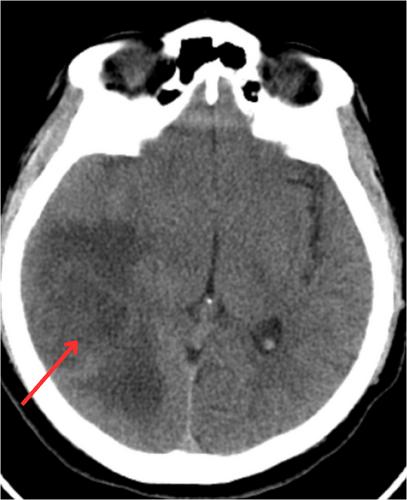
A 40-year-old male with no significant past medical history presented to the emergency department with a severe headache for 3 days. On physical examination, the patient appeared very uncomfortable and was hypoxic on room air to 86%. He was ambulatory with no apparent focal neurologic deficits, but his fingers were clubbed. Electrocardiogram showed normal sinus rhythm. Computed tomography (CT) angiogram of the chest incidentally revealed a portosystemic shunt in the left upper quadrant. CT brain and magnetic resonance imaging (MRI) brain with and without contrast were obtained (Figures 1 and 2).

图片结节和缺氧
一名 40 岁的男性因剧烈头痛 3 天来急诊就诊,既往无重大病史。经体格检查,患者看起来很不舒服,室内空气缺氧率高达 86%。他行动自如,没有明显的局灶性神经功能缺损,但手指呈棍棒状。心电图显示窦性心律正常。胸部计算机断层扫描(CT)血管造影偶然发现左上腹有一个门静脉分流。脑部 CT 和核磁共振成像(MRI)显示有造影剂和无造影剂(图 1 和图 2)。
本文章由计算机程序翻译,如有差异,请以英文原文为准。
求助全文
约1分钟内获得全文
求助全文
来源期刊
CiteScore
4.10
自引率
0.00%
发文量
0
审稿时长
5 weeks

 求助内容:
求助内容: 应助结果提醒方式:


