{"title":"Two-devices-in-one-channel method for minor papilla cannulation","authors":"Kiyoaki Ochi, Tsuneyoshi Ogawa, Toru Ueki","doi":"10.1111/den.14907","DOIUrl":null,"url":null,"abstract":"<p>Minor papilla cannulation is performed in patients with pancreas divisum and acute recurrent pancreatitis<span><sup>1</sup></span>; however, it can be a technically challenging procedure.<span><sup>2</sup></span> We demonstrated the two-devices-in-one-channel method for minor papilla cannulation. A 50-year-old man was admitted to our hospital for recurrent pancreatitis. Pancreas divisum was suspected to be the cause of the recurrent pancreatitis. Subsequently, the patient underwent endoscopic treatment, during which a duodenoscope (model TJF 290 V; Olympus, Tokyo, Japan) was advanced to the minor duodenal papilla. We initially attempted wire-guided cannulation; we were unsuccessful because of the small size of the minor papilla, its loose fixation, and susceptibility to respiratory variability (Fig. 1). Therefore, we attempted minor papilla cannulation using the two-devices-in-one-channel method (Video S1). A slim catheter (model PR-110Q; Olympus), loaded with a 0.025 inch guidewire (Radifocus; Terumo, Tokyo, Japan) and small biopsy forceps (Radial Jaw4P; Boston Scientific, Marlborough, MA, USA) were inserted into the same channel of the duodenoscope. The forceps were then used to grasp the anal side of the minor papilla and pull it towards the scope to retract the catheter tip into the minor papilla. Following this procedure, we fixed the minor papilla and aligned the catheter with the pancreatic duct axis. After successful cannulation, sphincterotomy was performed, followed by the placement of a 7F, 5 cm pancreatic stent (Advanix; Boston Scientific). During the wire-guided cannulation, the endoscopist pushed the cannula with force, which can cause the pancreatic duct axis to bend easily if the minor papilla is inadequately fixed. However, using the two-devices-in-one-channel method and pulling the minor papilla toward the scope can help adjust the axis of the catheter to the pancreatic duct as it straightens the bend in the pancreatic duct. This method is, therefore, an effective technique not only for biliary cannulation<span><sup>3, 4</sup></span> but also for minor papilla cannulation.</p><p>Authors declare no conflict of interest for this article.</p>","PeriodicalId":159,"journal":{"name":"Digestive Endoscopy","volume":"36 11","pages":"1290-1291"},"PeriodicalIF":5.0000,"publicationDate":"2024-08-27","publicationTypes":"Journal Article","fieldsOfStudy":null,"isOpenAccess":false,"openAccessPdf":"https://onlinelibrary.wiley.com/doi/epdf/10.1111/den.14907","citationCount":"0","resultStr":null,"platform":"Semanticscholar","paperid":null,"PeriodicalName":"Digestive Endoscopy","FirstCategoryId":"3","ListUrlMain":"https://onlinelibrary.wiley.com/doi/10.1111/den.14907","RegionNum":2,"RegionCategory":"医学","ArticlePicture":[],"TitleCN":null,"AbstractTextCN":null,"PMCID":null,"EPubDate":"","PubModel":"","JCR":"Q1","JCRName":"GASTROENTEROLOGY & HEPATOLOGY","Score":null,"Total":0}
引用次数: 0
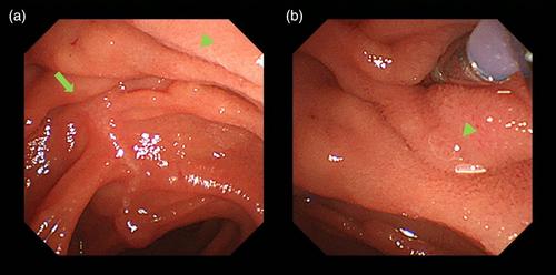
Abstract
Minor papilla cannulation is performed in patients with pancreas divisum and acute recurrent pancreatitis1; however, it can be a technically challenging procedure.2 We demonstrated the two-devices-in-one-channel method for minor papilla cannulation. A 50-year-old man was admitted to our hospital for recurrent pancreatitis. Pancreas divisum was suspected to be the cause of the recurrent pancreatitis. Subsequently, the patient underwent endoscopic treatment, during which a duodenoscope (model TJF 290 V; Olympus, Tokyo, Japan) was advanced to the minor duodenal papilla. We initially attempted wire-guided cannulation; we were unsuccessful because of the small size of the minor papilla, its loose fixation, and susceptibility to respiratory variability (Fig. 1). Therefore, we attempted minor papilla cannulation using the two-devices-in-one-channel method (Video S1). A slim catheter (model PR-110Q; Olympus), loaded with a 0.025 inch guidewire (Radifocus; Terumo, Tokyo, Japan) and small biopsy forceps (Radial Jaw4P; Boston Scientific, Marlborough, MA, USA) were inserted into the same channel of the duodenoscope. The forceps were then used to grasp the anal side of the minor papilla and pull it towards the scope to retract the catheter tip into the minor papilla. Following this procedure, we fixed the minor papilla and aligned the catheter with the pancreatic duct axis. After successful cannulation, sphincterotomy was performed, followed by the placement of a 7F, 5 cm pancreatic stent (Advanix; Boston Scientific). During the wire-guided cannulation, the endoscopist pushed the cannula with force, which can cause the pancreatic duct axis to bend easily if the minor papilla is inadequately fixed. However, using the two-devices-in-one-channel method and pulling the minor papilla toward the scope can help adjust the axis of the catheter to the pancreatic duct as it straightens the bend in the pancreatic duct. This method is, therefore, an effective technique not only for biliary cannulation3, 4 but also for minor papilla cannulation.
Authors declare no conflict of interest for this article.
期刊介绍:
Digestive Endoscopy (DEN) is the official journal of the Japan Gastroenterological Endoscopy Society, the Asian Pacific Society for Digestive Endoscopy and the World Endoscopy Organization. Digestive Endoscopy serves as a medium for presenting original articles that offer significant contributions to knowledge in the broad field of endoscopy. The Journal also includes Reviews, Original Articles, How I Do It, Case Reports (only of exceptional interest and novelty are accepted), Letters, Techniques and Images, abstracts and news items that may be of interest to endoscopists.

 求助内容:
求助内容: 应助结果提醒方式:


