Harald Krenzlin, Dominik M A Wesp, Anika A E Korinek, Henning Ubbens, Jakob Volland, Julia Masomi-Bornwasser, Katharina J Weber, Dominik Mole, Clemens Sommer, Florian Ringel, Beat Alessandri, Naureen Keric
{"title":"Effects of Argon in the Acute Phase of Subarachnoid Hemorrhage in an Endovascular Perforation Model in Rats.","authors":"Harald Krenzlin, Dominik M A Wesp, Anika A E Korinek, Henning Ubbens, Jakob Volland, Julia Masomi-Bornwasser, Katharina J Weber, Dominik Mole, Clemens Sommer, Florian Ringel, Beat Alessandri, Naureen Keric","doi":"10.1007/s12028-024-02090-3","DOIUrl":null,"url":null,"abstract":"<p><strong>Background: </strong>Subarachnoid hemorrhage (SAH) is a devastating disease with high morbidity and mortality. Neuroprotective effects of the noble gas argon have been shown in animal models of ischemia. The aim of this study was to investigate the effects of argon in the immediate early phase of SAH in a rat model.</p><p><strong>Methods: </strong>A total of 19 male Wistar rats were randomly assigned to three treatment groups. SAH was induced using a endovascular filament perforation model. Cerebral blood flow, mean arterial blood pressure (MAP), and body temperature were measured continuously. Group A received 2 h of ventilation by 50% argon/50% O<sub>2</sub> (n = 7) immediately following SAH. Group B underwent a sham operation and was also ventilated by 50% argon/50% O<sub>2</sub> (n = 6). Group C underwent SAH and 50% O<sub>2</sub>/50% N<sub>2</sub> ventilation (n = 6). Preoperative and postoperative neurological and behavioral testing were performed. Histology and immunohistochemistry were used to evaluate the extent of brain injury and vasospasm.</p><p><strong>Results: </strong>The cerebral blood flow dropped in both treatment groups after SAH induction (SAH, 63.0 ± 11.6% of baseline; SAH + argon, 80.2 ± 8.2% of baseline). During SAH, MAP increased (135.2 ± 10.5%) compared with baseline values (85.8 ± 26.0 mm Hg) and normalized thereafter. MAP in both groups showed no significant differences (p = 0.3123). Immunohistochemical staining for neuronal nuclear antigen demonstrated a decrease of hippocampal immunoreactivity after SAH in the cornu ammonis region (CA) 1-3 compared with baseline hippocampal immunoreactivity (p = 0.0127). Animals in the argon-ventilated group showed less neuronal loss compared with untreated SAH animals (p < 0.0001). Ionized calcium-binding adaptor molecule 1 staining showed a decreased accumulation after SAH + argon (CA1, 2.57 ± 2.35%; CA2, 1.89 ± 1.89%; CA3, 2.19 ± 1.99%; DG, 2.6 ± 2.24%) compared with untreated SAH animals (CA1, 5.48 ± 2.39%; CA2, 4.85 ± 4.06%; CA3, 4.22 ± 3.01%; dentate gyrus (DG), 3.82 ± 3.23%; p = 0.0007). The neuroscore assessment revealed no treatment benefit after SAH compared with baseline (p = 0.385).</p><p><strong>Conclusion: </strong>In the present study, neuroprotective effects of argon occurred early after SAH. Because neurological deterioration was similar in the preadministration and absence of argon, it remains uncertain if neuroprotective effects translate in improved outcome over time.</p>","PeriodicalId":19118,"journal":{"name":"Neurocritical Care","volume":" ","pages":"532-540"},"PeriodicalIF":3.1000,"publicationDate":"2025-04-01","publicationTypes":"Journal Article","fieldsOfStudy":null,"isOpenAccess":false,"openAccessPdf":"https://www.ncbi.nlm.nih.gov/pmc/articles/PMC11950149/pdf/","citationCount":"0","resultStr":null,"platform":"Semanticscholar","paperid":null,"PeriodicalName":"Neurocritical Care","FirstCategoryId":"3","ListUrlMain":"https://doi.org/10.1007/s12028-024-02090-3","RegionNum":3,"RegionCategory":"医学","ArticlePicture":[],"TitleCN":null,"AbstractTextCN":null,"PMCID":null,"EPubDate":"2024/8/22 0:00:00","PubModel":"Epub","JCR":"Q2","JCRName":"CLINICAL NEUROLOGY","Score":null,"Total":0}
引用次数: 0
Abstract
Background: Subarachnoid hemorrhage (SAH) is a devastating disease with high morbidity and mortality. Neuroprotective effects of the noble gas argon have been shown in animal models of ischemia. The aim of this study was to investigate the effects of argon in the immediate early phase of SAH in a rat model.
Methods: A total of 19 male Wistar rats were randomly assigned to three treatment groups. SAH was induced using a endovascular filament perforation model. Cerebral blood flow, mean arterial blood pressure (MAP), and body temperature were measured continuously. Group A received 2 h of ventilation by 50% argon/50% O2 (n = 7) immediately following SAH. Group B underwent a sham operation and was also ventilated by 50% argon/50% O2 (n = 6). Group C underwent SAH and 50% O2/50% N2 ventilation (n = 6). Preoperative and postoperative neurological and behavioral testing were performed. Histology and immunohistochemistry were used to evaluate the extent of brain injury and vasospasm.
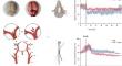
Results: The cerebral blood flow dropped in both treatment groups after SAH induction (SAH, 63.0 ± 11.6% of baseline; SAH + argon, 80.2 ± 8.2% of baseline). During SAH, MAP increased (135.2 ± 10.5%) compared with baseline values (85.8 ± 26.0 mm Hg) and normalized thereafter. MAP in both groups showed no significant differences (p = 0.3123). Immunohistochemical staining for neuronal nuclear antigen demonstrated a decrease of hippocampal immunoreactivity after SAH in the cornu ammonis region (CA) 1-3 compared with baseline hippocampal immunoreactivity (p = 0.0127). Animals in the argon-ventilated group showed less neuronal loss compared with untreated SAH animals (p < 0.0001). Ionized calcium-binding adaptor molecule 1 staining showed a decreased accumulation after SAH + argon (CA1, 2.57 ± 2.35%; CA2, 1.89 ± 1.89%; CA3, 2.19 ± 1.99%; DG, 2.6 ± 2.24%) compared with untreated SAH animals (CA1, 5.48 ± 2.39%; CA2, 4.85 ± 4.06%; CA3, 4.22 ± 3.01%; dentate gyrus (DG), 3.82 ± 3.23%; p = 0.0007). The neuroscore assessment revealed no treatment benefit after SAH compared with baseline (p = 0.385).
Conclusion: In the present study, neuroprotective effects of argon occurred early after SAH. Because neurological deterioration was similar in the preadministration and absence of argon, it remains uncertain if neuroprotective effects translate in improved outcome over time.
期刊介绍:
Neurocritical Care is a peer reviewed scientific publication whose major goal is to disseminate new knowledge on all aspects of acute neurological care. It is directed towards neurosurgeons, neuro-intensivists, neurologists, anesthesiologists, emergency physicians, and critical care nurses treating patients with urgent neurologic disorders. These are conditions that may potentially evolve rapidly and could need immediate medical or surgical intervention. Neurocritical Care provides a comprehensive overview of current developments in intensive care neurology, neurosurgery and neuroanesthesia and includes information about new therapeutic avenues and technological innovations. Neurocritical Care is the official journal of the Neurocritical Care Society.
 求助内容:
求助内容: 应助结果提醒方式:


