Potential mechanisms of cancer stem-like progenitor T-cell bio-behaviours

IF 7.9 1区 医学 Q1 MEDICINE, RESEARCH & EXPERIMENTAL
Ling Ni
{"title":"Potential mechanisms of cancer stem-like progenitor T-cell bio-behaviours","authors":"Ling Ni","doi":"10.1002/ctm2.1817","DOIUrl":null,"url":null,"abstract":"<div>\n \n \n <section>\n \n <p>In situations involving continuous exposure to antigens, such as chronic infections or cancer, antigen-specific CD8<sup>+</sup> T cells can become dysfunctional or exhausted. This change is marked by increased expression levels of inhibitory receptors (PD-1 and Tim-3). Stem-like progenitor exhausted (Tpex) cells, a subset of exhausted cells that express TCF-1 and are mainly found in the lymph nodes, demonstrate the ability to self-renew and exhibit a high rate of proliferation. Tpex cells can further differentiate into transitional intermediate exhausted (Tex-int) cells and terminally exhausted (Tex-term) cells. Alternatively, they can directly differentiate into Tex-term cells. Tpex cells are the predominant subset that respond to immune checkpoint inhibitors (ICI), making them a prime candidate for improving the efficacy of ICI therapy. This review article aimed to present the latest developments in the field of Tpex formation, expansion, and differentiation in the context of cancer, as well as their responses to ICIs in cancer immunotherapy. Consequently, it may be possible to develop novel treatments that exclusively target Tpex cells, thus improving overall treatment outcomes.</p>\n </section>\n \n <section>\n \n <h3> Key points</h3>\n \n <div>\n <ul>\n \n <li>\n <p>Tpex cells are located in lymph nodes and TLS.</p>\n </li>\n \n <li>\n <p>Several pathways control the differentiation trajectories of Tpex cells, including epigenetic factors, transcription factors, cytokines, age, sex, etc.</p>\n </li>\n </ul>\n </div>\n </section>\n </div>","PeriodicalId":10189,"journal":{"name":"Clinical and Translational Medicine","volume":"14 8","pages":""},"PeriodicalIF":7.9000,"publicationDate":"2024-08-21","publicationTypes":"Journal Article","fieldsOfStudy":null,"isOpenAccess":false,"openAccessPdf":"https://onlinelibrary.wiley.com/doi/epdf/10.1002/ctm2.1817","citationCount":"0","resultStr":null,"platform":"Semanticscholar","paperid":null,"PeriodicalName":"Clinical and Translational Medicine","FirstCategoryId":"3","ListUrlMain":"https://onlinelibrary.wiley.com/doi/10.1002/ctm2.1817","RegionNum":1,"RegionCategory":"医学","ArticlePicture":[],"TitleCN":null,"AbstractTextCN":null,"PMCID":null,"EPubDate":"","PubModel":"","JCR":"Q1","JCRName":"MEDICINE, RESEARCH & EXPERIMENTAL","Score":null,"Total":0}
引用次数: 0

Abstract

In situations involving continuous exposure to antigens, such as chronic infections or cancer, antigen-specific CD8+ T cells can become dysfunctional or exhausted. This change is marked by increased expression levels of inhibitory receptors (PD-1 and Tim-3). Stem-like progenitor exhausted (Tpex) cells, a subset of exhausted cells that express TCF-1 and are mainly found in the lymph nodes, demonstrate the ability to self-renew and exhibit a high rate of proliferation. Tpex cells can further differentiate into transitional intermediate exhausted (Tex-int) cells and terminally exhausted (Tex-term) cells. Alternatively, they can directly differentiate into Tex-term cells. Tpex cells are the predominant subset that respond to immune checkpoint inhibitors (ICI), making them a prime candidate for improving the efficacy of ICI therapy. This review article aimed to present the latest developments in the field of Tpex formation, expansion, and differentiation in the context of cancer, as well as their responses to ICIs in cancer immunotherapy. Consequently, it may be possible to develop novel treatments that exclusively target Tpex cells, thus improving overall treatment outcomes.

Key points

  • Tpex cells are located in lymph nodes and TLS.

  • Several pathways control the differentiation trajectories of Tpex cells, including epigenetic factors, transcription factors, cytokines, age, sex, etc.
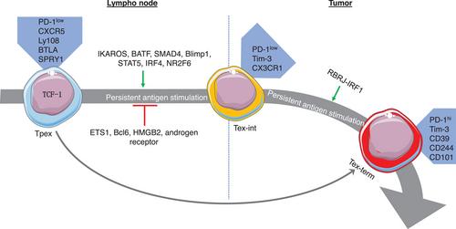
Abstract Image

癌症干样祖细胞 T 细胞生物行为的潜在机制。
在持续暴露于抗原(如慢性感染或癌症)的情况下,抗原特异性 CD8+ T 细胞会出现功能障碍或衰竭。这种变化的特征是抑制性受体(PD-1 和 Tim-3)的表达水平升高。干样祖细胞衰竭(Tpex)细胞是衰竭细胞的一个亚群,表达 TCF-1,主要存在于淋巴结中。Tpex细胞可进一步分化为过渡中间衰竭(Tex-int)细胞和终末衰竭(Tex-term)细胞。或者,它们也可以直接分化成 Tex-term 细胞。Tpex细胞是对免疫检查点抑制剂(ICI)有反应的主要亚群,是提高ICI疗效的主要候选细胞。这篇综述文章旨在介绍癌症背景下 Tpex 形成、扩增和分化领域的最新进展,以及它们在癌症免疫疗法中对 ICIs 的反应。因此,有可能开发出专门针对 Tpex 细胞的新型疗法,从而改善整体治疗效果。要点Tpex细胞位于淋巴结和TLS中。Tpex细胞的分化轨迹受多种途径控制,包括表观遗传因子、转录因子、细胞因子、年龄、性别等。
本文章由计算机程序翻译,如有差异,请以英文原文为准。
求助全文
约1分钟内获得全文 求助全文
来源期刊
CiteScore
15.90
自引率
1.90%
发文量
450
审稿时长
4 weeks
期刊介绍: Clinical and Translational Medicine (CTM) is an international, peer-reviewed, open-access journal dedicated to accelerating the translation of preclinical research into clinical applications and fostering communication between basic and clinical scientists. It highlights the clinical potential and application of various fields including biotechnologies, biomaterials, bioengineering, biomarkers, molecular medicine, omics science, bioinformatics, immunology, molecular imaging, drug discovery, regulation, and health policy. With a focus on the bench-to-bedside approach, CTM prioritizes studies and clinical observations that generate hypotheses relevant to patients and diseases, guiding investigations in cellular and molecular medicine. The journal encourages submissions from clinicians, researchers, policymakers, and industry professionals.
×
引用
GB/T 7714-2015
复制
MLA
复制
APA
复制
导出至
BibTeX EndNote RefMan NoteFirst NoteExpress
×
提示
您的信息不完整,为了账户安全,请先补充。
现在去补充
×
提示
您因"违规操作"
具体请查看互助需知
我知道了
×
提示
确定
请完成安全验证×
copy
已复制链接
快去分享给好友吧!
我知道了
右上角分享
点击右上角分享
0
联系我们:info@booksci.cn Book学术提供免费学术资源搜索服务,方便国内外学者检索中英文文献。致力于提供最便捷和优质的服务体验。 Copyright © 2023 布克学术 All rights reserved.
京ICP备2023020795号-1
ghs 京公网安备 11010802042870号
Book学术文献互助
Book学术文献互助群
群 号:481959085
Book学术官方微信