{"title":"Conserved N-terminal Regulation of the ACA8 Calcium Pump with Two Calmodulin Binding Sites","authors":"","doi":"10.1016/j.jmb.2024.168747","DOIUrl":null,"url":null,"abstract":"<div><p>The autoinhibited plasma membrane calcium ATPase ACA8 from <em>A. thaliana</em> has an N-terminal autoinhibitory domain. Binding of calcium-loaded calmodulin at two sites located at residues 42–62 and 74–96 relieves autoinhibition of ACA8 activity.</p><p>Through activity studies and a yeast complementation assay we investigated wild-type (WT) and N-terminally truncated ACA8 constructs (Δ20, Δ30, Δ35, Δ37, Δ40, Δ74 and Δ100) to explore the role of conserved motifs in the N-terminal segment preceding the calmodulin binding sites. Furthermore, we purified WT, Δ20- and Δ100-ACA8, tested activity <em>in vitro</em> and performed structural studies of purified Δ20-ACA8 stabilized in a lipid nanodisc to explore the mechanism of autoinhibition.</p><p>We show that an N-terminal segment between residues 20 and 35 including conserved Phe32, upstream of the calmodulin binding sites, is important for autoinhibition and the activation by calmodulin. Cryo-EM structure determination at 3.3 Å resolution of a beryllium fluoride inhibited E2 form, and at low resolution for an E1 state combined with AlphaFold prediction provide a model for autoinhibition, consistent with the mutational studies.</p></div>","PeriodicalId":369,"journal":{"name":"Journal of Molecular Biology","volume":null,"pages":null},"PeriodicalIF":4.7000,"publicationDate":"2024-08-20","publicationTypes":"Journal Article","fieldsOfStudy":null,"isOpenAccess":false,"openAccessPdf":"https://www.sciencedirect.com/science/article/pii/S0022283624003565/pdfft?md5=b8cc8fcfc2d15016e9e97d1a6090aedc&pid=1-s2.0-S0022283624003565-main.pdf","citationCount":"0","resultStr":null,"platform":"Semanticscholar","paperid":null,"PeriodicalName":"Journal of Molecular Biology","FirstCategoryId":"99","ListUrlMain":"https://www.sciencedirect.com/science/article/pii/S0022283624003565","RegionNum":2,"RegionCategory":"生物学","ArticlePicture":[],"TitleCN":null,"AbstractTextCN":null,"PMCID":null,"EPubDate":"","PubModel":"","JCR":"Q1","JCRName":"BIOCHEMISTRY & MOLECULAR BIOLOGY","Score":null,"Total":0}
引用次数: 0
Abstract
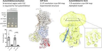
The autoinhibited plasma membrane calcium ATPase ACA8 from A. thaliana has an N-terminal autoinhibitory domain. Binding of calcium-loaded calmodulin at two sites located at residues 42–62 and 74–96 relieves autoinhibition of ACA8 activity.
Through activity studies and a yeast complementation assay we investigated wild-type (WT) and N-terminally truncated ACA8 constructs (Δ20, Δ30, Δ35, Δ37, Δ40, Δ74 and Δ100) to explore the role of conserved motifs in the N-terminal segment preceding the calmodulin binding sites. Furthermore, we purified WT, Δ20- and Δ100-ACA8, tested activity in vitro and performed structural studies of purified Δ20-ACA8 stabilized in a lipid nanodisc to explore the mechanism of autoinhibition.
We show that an N-terminal segment between residues 20 and 35 including conserved Phe32, upstream of the calmodulin binding sites, is important for autoinhibition and the activation by calmodulin. Cryo-EM structure determination at 3.3 Å resolution of a beryllium fluoride inhibited E2 form, and at low resolution for an E1 state combined with AlphaFold prediction provide a model for autoinhibition, consistent with the mutational studies.
期刊介绍:
Journal of Molecular Biology (JMB) provides high quality, comprehensive and broad coverage in all areas of molecular biology. The journal publishes original scientific research papers that provide mechanistic and functional insights and report a significant advance to the field. The journal encourages the submission of multidisciplinary studies that use complementary experimental and computational approaches to address challenging biological questions.
Research areas include but are not limited to: Biomolecular interactions, signaling networks, systems biology; Cell cycle, cell growth, cell differentiation; Cell death, autophagy; Cell signaling and regulation; Chemical biology; Computational biology, in combination with experimental studies; DNA replication, repair, and recombination; Development, regenerative biology, mechanistic and functional studies of stem cells; Epigenetics, chromatin structure and function; Gene expression; Membrane processes, cell surface proteins and cell-cell interactions; Methodological advances, both experimental and theoretical, including databases; Microbiology, virology, and interactions with the host or environment; Microbiota mechanistic and functional studies; Nuclear organization; Post-translational modifications, proteomics; Processing and function of biologically important macromolecules and complexes; Molecular basis of disease; RNA processing, structure and functions of non-coding RNAs, transcription; Sorting, spatiotemporal organization, trafficking; Structural biology; Synthetic biology; Translation, protein folding, chaperones, protein degradation and quality control.
 求助内容:
求助内容: 应助结果提醒方式:


