Multiparametric-magnetic resonance imaging (mp-MRI) of the prostate and Urolift: Identifying artefact size, location and clinical implications

IF 1.6 Q3 UROLOGY & NEPHROLOGY
BJUI compass Pub Date : 2024-06-01 DOI:10.1002/bco2.392
Cameron James Parkin, Rajeev Jyoti, Peter Chin
{"title":"Multiparametric-magnetic resonance imaging (mp-MRI) of the prostate and Urolift: Identifying artefact size, location and clinical implications","authors":"Cameron James Parkin,&nbsp;Rajeev Jyoti,&nbsp;Peter Chin","doi":"10.1002/bco2.392","DOIUrl":null,"url":null,"abstract":"<div>\n \n \n <section>\n \n <h3> Objectives</h3>\n \n <p>We sought to define the degree of artefact caused by prostatic urethral lift (PUL) on multiparametric-magnetic resonance imaging (mp-MRI) to determine the location, size of artefact and if the device could potentially obscure a diagnosis of prostate cancer.</p>\n </section>\n \n <section>\n \n <h3> Methods</h3>\n \n <p>Ten patients were prospectively enrolled to undergo PUL for treatment of benign prostatic hyperplasia and follow-up imaging. A standard mp-MRI protocol using a 3.0 Tesla scanner was performed prior to and following Urolift insertion. Pre- and post-PUL images were compared to measure maximum artefact diameter around each implant in each MRI parameter. A transverse relaxation time weighted (T2) artefact reduction protocol was also evaluated. The location of each artefact was then compared to a separate database of 225 consecutive patients who underwent magnetic resonance guided prostate biopsies.</p>\n </section>\n \n <section>\n \n <h3> Results</h3>\n \n <p>Artefact occurred around the stainless steel urethral implant component only. Mean T2 artefact maximum diameter was 7.7 mm (sd = 1.71 mm), with an artefact reduction protocol reducing this to 5.4 mm (sd = 1.43). Mean dynamic-contrast-enhancement artefact was 10 mm (sd = 2.5 mm), and mean diffusion-weighted-imaging artefact was 28.2 mm (sd = 7.8 mm). All artefacts were confined to the posterior transition zone only. In the 225 consecutive patients who had undergone magnetic resonance guided prostate biopsies, there were 55 positive biopsies with prostate cancer, with 13 cases found in the transition zones and no cancer identified solely in the posterior transitional zone.</p>\n </section>\n \n <section>\n \n <h3> Conclusions</h3>\n \n <p>The stainless steel urethral component of the PUL does cause artefact, which is confined to the posterior transition zone only. PUL artefact occurs in an area of the prostate that has a very low incidence of a single focus of prostate cancer. If there is concern for prostate cancer in the posterior TZ (e.g. if every other area is clear with a high PSA), this area can undergo targeted biopsy.</p>\n </section>\n </div>","PeriodicalId":72420,"journal":{"name":"BJUI compass","volume":"5 8","pages":"770-775"},"PeriodicalIF":1.6000,"publicationDate":"2024-06-01","publicationTypes":"Journal Article","fieldsOfStudy":null,"isOpenAccess":false,"openAccessPdf":"https://onlinelibrary.wiley.com/doi/epdf/10.1002/bco2.392","citationCount":"0","resultStr":null,"platform":"Semanticscholar","paperid":null,"PeriodicalName":"BJUI compass","FirstCategoryId":"1085","ListUrlMain":"https://onlinelibrary.wiley.com/doi/10.1002/bco2.392","RegionNum":0,"RegionCategory":null,"ArticlePicture":[],"TitleCN":null,"AbstractTextCN":null,"PMCID":null,"EPubDate":"","PubModel":"","JCR":"Q3","JCRName":"UROLOGY & NEPHROLOGY","Score":null,"Total":0}
引用次数: 0

Abstract

Objectives

We sought to define the degree of artefact caused by prostatic urethral lift (PUL) on multiparametric-magnetic resonance imaging (mp-MRI) to determine the location, size of artefact and if the device could potentially obscure a diagnosis of prostate cancer.

Methods

Ten patients were prospectively enrolled to undergo PUL for treatment of benign prostatic hyperplasia and follow-up imaging. A standard mp-MRI protocol using a 3.0 Tesla scanner was performed prior to and following Urolift insertion. Pre- and post-PUL images were compared to measure maximum artefact diameter around each implant in each MRI parameter. A transverse relaxation time weighted (T2) artefact reduction protocol was also evaluated. The location of each artefact was then compared to a separate database of 225 consecutive patients who underwent magnetic resonance guided prostate biopsies.

Results

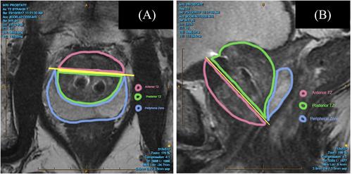
Artefact occurred around the stainless steel urethral implant component only. Mean T2 artefact maximum diameter was 7.7 mm (sd = 1.71 mm), with an artefact reduction protocol reducing this to 5.4 mm (sd = 1.43). Mean dynamic-contrast-enhancement artefact was 10 mm (sd = 2.5 mm), and mean diffusion-weighted-imaging artefact was 28.2 mm (sd = 7.8 mm). All artefacts were confined to the posterior transition zone only. In the 225 consecutive patients who had undergone magnetic resonance guided prostate biopsies, there were 55 positive biopsies with prostate cancer, with 13 cases found in the transition zones and no cancer identified solely in the posterior transitional zone.

Conclusions

The stainless steel urethral component of the PUL does cause artefact, which is confined to the posterior transition zone only. PUL artefact occurs in an area of the prostate that has a very low incidence of a single focus of prostate cancer. If there is concern for prostate cancer in the posterior TZ (e.g. if every other area is clear with a high PSA), this area can undergo targeted biopsy.
Abstract Image

前列腺多参数磁共振成像(mp-MRI)和尿道前列腺电切术:识别伪影大小、位置和临床影响
目的 我们试图定义前列腺尿道提升器(PUL)在多参数磁共振成像(mp-MRI)上造成的伪影程度,以确定伪影的位置、大小以及该装置是否有可能掩盖前列腺癌的诊断。 方法 前瞻性地招募了十名患者,让他们接受 PUL 治疗良性前列腺增生并进行后续成像。在插入 Urolift 之前和之后,使用 3.0 特斯拉扫描仪执行了标准 mp-MRI 方案。对插入前和插入后的图像进行比较,以测量每个磁共振成像参数中每个植入物周围的最大伪影直径。此外,还评估了横向弛豫时间加权(T2)伪影减少方案。然后将每个伪影的位置与磁共振引导下进行前列腺活检的 225 名连续患者的单独数据库进行比较。 结果 伪影仅出现在不锈钢尿道植入组件周围。平均 T2 伪影最大直径为 7.7 毫米(sd = 1.71 毫米),伪影减少方案将其减少到 5.4 毫米(sd = 1.43)。动态对比度增强的平均伪影为 10 毫米(sd = 2.5 毫米),扩散加权成像的平均伪影为 28.2 毫米(sd = 7.8 毫米)。所有的伪影都仅限于后过渡区。在连续接受磁共振引导前列腺活检的 225 名患者中,有 55 例前列腺癌活检阳性病例,其中 13 例发现于过渡区,没有发现仅位于后过渡区的癌症。 结论 PUL 的不锈钢尿道组件确实会造成伪影,但仅局限于后过渡区。PUL 伪影发生在前列腺中单个前列腺癌灶发生率极低的区域。如果担心后过渡区会出现前列腺癌(例如,如果其他区域都很清晰,但 PSA 很高),可以对该区域进行靶向活检。
本文章由计算机程序翻译,如有差异,请以英文原文为准。
求助全文
约1分钟内获得全文 求助全文
来源期刊
CiteScore
2.30
自引率
0.00%
发文量
0
审稿时长
12 weeks
×
引用
GB/T 7714-2015
复制
MLA
复制
APA
复制
导出至
BibTeX EndNote RefMan NoteFirst NoteExpress
×
提示
您的信息不完整,为了账户安全,请先补充。
现在去补充
×
提示
您因"违规操作"
具体请查看互助需知
我知道了
×
提示
确定
请完成安全验证×
copy
已复制链接
快去分享给好友吧!
我知道了
右上角分享
点击右上角分享
0
联系我们:info@booksci.cn Book学术提供免费学术资源搜索服务,方便国内外学者检索中英文文献。致力于提供最便捷和优质的服务体验。 Copyright © 2023 布克学术 All rights reserved.
京ICP备2023020795号-1
ghs 京公网安备 11010802042870号
Book学术文献互助
Book学术文献互助群
群 号:481959085
Book学术官方微信