{"title":"Inter-tissue differences in oxidative stress susceptibility reveal a less stable endothelial barrier in the brain than in the retina","authors":"","doi":"10.1016/j.expneurol.2024.114919","DOIUrl":null,"url":null,"abstract":"<div><p>Oxidative stress can impair the endothelial barrier and thereby enable autoantibody migration in Neuromyelitis optica spectrum disorder (NMOSD). Tissue-specific vulnerability to autoantibody-mediated damage could be explained by a differential, tissue-dependent endothelial susceptibility to oxidative stress. In this study, we aim to investigate the barrier integrity and complement profiles of brain and retinal endothelial cells under oxygen-induced oxidative stress to address the question of whether the pathomechanism of NMOSD preferentially affects the brain or the retina.</p><p>Primary human brain microvascular endothelial cells (HBMEC) and primary human retinal endothelial cells (HREC) were cultivated at different cell densities (2.5*10<sup>4</sup> to 2*10<sup>5</sup> cells/cm<sup>2</sup>) for real-time cell analysis. Both cell types were exposed to 100, 500 and 2500 μM H<sub>2</sub>O<sub>2</sub>. Immunostaining (CD31, VE-cadherin, ZO-1) and Western blot, as well as complement protein secretion using multiplex ELISA were performed.</p><p>HBMEC and HREC cell growth phases were cell type-specific. While HBMEC cell growth could be categorized into an initial peak, proliferation phase, plateau phase, and barrier breakdown phase, HREC showed no proliferation phase, but entered the plateau phase immediately after an initial peak. The plateau phase was 7 h shorter in HREC. Both cell types displayed a short-term, dose-dependent adaptive response to H<sub>2</sub>O<sub>2</sub>. Remarkably, at 100 μM H<sub>2</sub>O<sub>2</sub>, the transcellular resistance of HBMEC exceeded that of untreated cells. 500 μM H<sub>2</sub>O<sub>2</sub> exerted a more disruptive effect on the HBMEC transcellular resistance than on HREC. Both cell types secreted complement factors H (FH) and I (FI), with FH secretion remaining stable after 2 h, but FI secretion decreasing at higher H<sub>2</sub>O<sub>2</sub> concentrations.</p><p>The observed differences in resistance to oxidative stress between primary brain and retinal endothelial cells may have implications for further studies of NMOSD and other autoimmune diseases affecting the eye and brain. These findings may open novel perspectives for the understanding and treatment of such diseases.</p></div>","PeriodicalId":12246,"journal":{"name":"Experimental Neurology","volume":null,"pages":null},"PeriodicalIF":4.6000,"publicationDate":"2024-08-12","publicationTypes":"Journal Article","fieldsOfStudy":null,"isOpenAccess":false,"openAccessPdf":"https://www.sciencedirect.com/science/article/pii/S0014488624002450/pdfft?md5=e836b6cea3f6c77b7ea0b41af746d83b&pid=1-s2.0-S0014488624002450-main.pdf","citationCount":"0","resultStr":null,"platform":"Semanticscholar","paperid":null,"PeriodicalName":"Experimental Neurology","FirstCategoryId":"3","ListUrlMain":"https://www.sciencedirect.com/science/article/pii/S0014488624002450","RegionNum":2,"RegionCategory":"医学","ArticlePicture":[],"TitleCN":null,"AbstractTextCN":null,"PMCID":null,"EPubDate":"","PubModel":"","JCR":"Q1","JCRName":"NEUROSCIENCES","Score":null,"Total":0}
引用次数: 0
Abstract
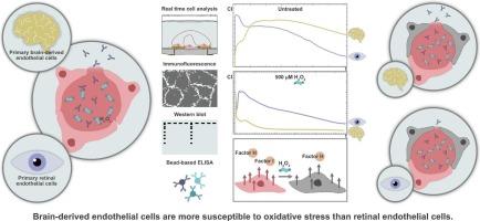
Oxidative stress can impair the endothelial barrier and thereby enable autoantibody migration in Neuromyelitis optica spectrum disorder (NMOSD). Tissue-specific vulnerability to autoantibody-mediated damage could be explained by a differential, tissue-dependent endothelial susceptibility to oxidative stress. In this study, we aim to investigate the barrier integrity and complement profiles of brain and retinal endothelial cells under oxygen-induced oxidative stress to address the question of whether the pathomechanism of NMOSD preferentially affects the brain or the retina.
Primary human brain microvascular endothelial cells (HBMEC) and primary human retinal endothelial cells (HREC) were cultivated at different cell densities (2.5*104 to 2*105 cells/cm2) for real-time cell analysis. Both cell types were exposed to 100, 500 and 2500 μM H2O2. Immunostaining (CD31, VE-cadherin, ZO-1) and Western blot, as well as complement protein secretion using multiplex ELISA were performed.
HBMEC and HREC cell growth phases were cell type-specific. While HBMEC cell growth could be categorized into an initial peak, proliferation phase, plateau phase, and barrier breakdown phase, HREC showed no proliferation phase, but entered the plateau phase immediately after an initial peak. The plateau phase was 7 h shorter in HREC. Both cell types displayed a short-term, dose-dependent adaptive response to H2O2. Remarkably, at 100 μM H2O2, the transcellular resistance of HBMEC exceeded that of untreated cells. 500 μM H2O2 exerted a more disruptive effect on the HBMEC transcellular resistance than on HREC. Both cell types secreted complement factors H (FH) and I (FI), with FH secretion remaining stable after 2 h, but FI secretion decreasing at higher H2O2 concentrations.
The observed differences in resistance to oxidative stress between primary brain and retinal endothelial cells may have implications for further studies of NMOSD and other autoimmune diseases affecting the eye and brain. These findings may open novel perspectives for the understanding and treatment of such diseases.
期刊介绍:
Experimental Neurology, a Journal of Neuroscience Research, publishes original research in neuroscience with a particular emphasis on novel findings in neural development, regeneration, plasticity and transplantation. The journal has focused on research concerning basic mechanisms underlying neurological disorders.
 求助内容:
求助内容: 应助结果提醒方式:


