Ferran Brugada-Bellsolà, Pilar Teixidor Rodríguez, Antonio González-Crespo, Sebastián Menéndez-Girón, Cristina Hostalot Panisello, Roser Garcia-Armengol, Carlos J Domínguez Alonso
{"title":"Intraoperative ultrasound and magnetic resonance comparative analysis in brain tumor surgery: a valuable tool to flatten ultrasound's learning curve.","authors":"Ferran Brugada-Bellsolà, Pilar Teixidor Rodríguez, Antonio González-Crespo, Sebastián Menéndez-Girón, Cristina Hostalot Panisello, Roser Garcia-Armengol, Carlos J Domínguez Alonso","doi":"10.1007/s00701-024-06228-2","DOIUrl":null,"url":null,"abstract":"<p><strong>Background: </strong>Intraoperative ultrasound (IOUS) is a profitable tool for neurosurgical procedures' assistance, especially in neuro-oncology. It is a rapid, ergonomic and reproducible technique. However, its known handicap is a steep learning curve for neurosurgeons. Here, we describe an interesting postoperative analysis that provides extra feedback after surgery, accelerating the learning process.</p><p><strong>Method: </strong>We conducted a descriptive retrospective unicenter study including patients operated from intra-axial brain tumors using neuronavigation (Curve, Brainlab) and IOUS (BK-5000, BK medical) guidance. All patients had preoperative Magnetic Resonance Imaging (MRI) prior to tumor resection. During surgery, 3D neuronavigated IOUS studies (n3DUS) were obtained through craniotomy N13C5 transducer's integration to the neuronavigation system. At least two n3DUS studies were obtained: prior to tumor resection and at the resection conclusion. A postoperative MRI was performed within 48 h. MRI and n3DUS studies were posteriorly fused and analyzed with Elements (Brainlab) planning software, permitting two comparative analyses: preoperative MRI compared to pre-resection n3DUS and postoperative MRI to post-resection n3DUS. Cases with incomplete MRI or n3DUS studies were withdrawn from the study.</p><p><strong>Results: </strong>From April 2022 to March 2024, 73 patients were operated assisted by IOUS. From them, 39 were included in the study. Analyses comparing preoperative MRI and pre-resection n3DUS showed great concordance of tumor volume (p < 0,001) between both modalities. Analysis comparing postoperative MRI and post-resection n3DUS also showed good concordance in residual tumor volume (RTV) in cases where gross total resection (GTR) was not achieved (p < 0,001). In two cases, RTV detected on MRI that was not detected intra-operatively with IOUS could be reviewed in detail to recheck its appearance.</p><p><strong>Conclusions: </strong>Post-operative comparative analyses between IOUS and MRI is a valuable tool for novel ultrasound users, as it enhances the amount of feedback provided by cases and could accelerate the learning process, flattening this technique's learning curve.</p>","PeriodicalId":7370,"journal":{"name":"Acta Neurochirurgica","volume":null,"pages":null},"PeriodicalIF":1.9000,"publicationDate":"2024-08-14","publicationTypes":"Journal Article","fieldsOfStudy":null,"isOpenAccess":false,"openAccessPdf":"","citationCount":"0","resultStr":null,"platform":"Semanticscholar","paperid":null,"PeriodicalName":"Acta Neurochirurgica","FirstCategoryId":"3","ListUrlMain":"https://doi.org/10.1007/s00701-024-06228-2","RegionNum":3,"RegionCategory":"医学","ArticlePicture":[],"TitleCN":null,"AbstractTextCN":null,"PMCID":null,"EPubDate":"","PubModel":"","JCR":"Q3","JCRName":"CLINICAL NEUROLOGY","Score":null,"Total":0}
引用次数: 0
Abstract
Background: Intraoperative ultrasound (IOUS) is a profitable tool for neurosurgical procedures' assistance, especially in neuro-oncology. It is a rapid, ergonomic and reproducible technique. However, its known handicap is a steep learning curve for neurosurgeons. Here, we describe an interesting postoperative analysis that provides extra feedback after surgery, accelerating the learning process.
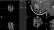
Method: We conducted a descriptive retrospective unicenter study including patients operated from intra-axial brain tumors using neuronavigation (Curve, Brainlab) and IOUS (BK-5000, BK medical) guidance. All patients had preoperative Magnetic Resonance Imaging (MRI) prior to tumor resection. During surgery, 3D neuronavigated IOUS studies (n3DUS) were obtained through craniotomy N13C5 transducer's integration to the neuronavigation system. At least two n3DUS studies were obtained: prior to tumor resection and at the resection conclusion. A postoperative MRI was performed within 48 h. MRI and n3DUS studies were posteriorly fused and analyzed with Elements (Brainlab) planning software, permitting two comparative analyses: preoperative MRI compared to pre-resection n3DUS and postoperative MRI to post-resection n3DUS. Cases with incomplete MRI or n3DUS studies were withdrawn from the study.
Results: From April 2022 to March 2024, 73 patients were operated assisted by IOUS. From them, 39 were included in the study. Analyses comparing preoperative MRI and pre-resection n3DUS showed great concordance of tumor volume (p < 0,001) between both modalities. Analysis comparing postoperative MRI and post-resection n3DUS also showed good concordance in residual tumor volume (RTV) in cases where gross total resection (GTR) was not achieved (p < 0,001). In two cases, RTV detected on MRI that was not detected intra-operatively with IOUS could be reviewed in detail to recheck its appearance.
Conclusions: Post-operative comparative analyses between IOUS and MRI is a valuable tool for novel ultrasound users, as it enhances the amount of feedback provided by cases and could accelerate the learning process, flattening this technique's learning curve.
期刊介绍:
The journal "Acta Neurochirurgica" publishes only original papers useful both to research and clinical work. Papers should deal with clinical neurosurgery - diagnosis and diagnostic techniques, operative surgery and results, postoperative treatment - or with research work in neuroscience if the underlying questions or the results are of neurosurgical interest. Reports on congresses are given in brief accounts. As official organ of the European Association of Neurosurgical Societies the journal publishes all announcements of the E.A.N.S. and reports on the activities of its member societies. Only contributions written in English will be accepted.

 求助内容:
求助内容: 应助结果提醒方式:


