{"title":"Advantages of SiPM-based digital PET/CT technology in nuclear medicine clinical practice: a systematic review—Part 1 oncological setting","authors":"Guido Rovera, Luca Urso, Federica Stracuzzi, Riccardo Laudicella, Viviana Frantellizzi, Chiara Cottignoli, Maria Gazzilli, Priscilla Guglielmo, Stefano Panareo, Laura Evangelista, Angelina Filice, Luca Burroni","doi":"10.1007/s40336-024-00653-0","DOIUrl":null,"url":null,"abstract":"<h3 data-test=\"abstract-sub-heading\">Purpose</h3><p>New-generation fully-digital PET/CT (dPET) scanners offer several technical advantages compared to analog (aPET) systems. This review aimed to summarize the current literature evidence about dPET technology clinical advantages.</p><h3 data-test=\"abstract-sub-heading\">Methods</h3><p>A systematic literature search of PubMed/MEDLINE and Embase databases was performed following PRISMA guidelines. The full-text articles methodological quality was independently assessed by four authors using the CASP-diagnostic study checklist.</p><h3 data-test=\"abstract-sub-heading\">Results</h3><p>Out of 510 articles, 81 were selected of which 42 related to oncology. In early-recurrent prostate cancer (PSA range ≤ 0.5 and 0.5–2.0 ng/ml), PSMA-dPET has shown a significantly higher detection rate compared to aPET especially for smaller lesions. A higher image quality and lesion detectability was reported in [<sup>18</sup>F]FDG studies on lung cancer and on mixed oncological cohorts, where metabolic TNM upstaging occurred in up to 32% of cases compared to aPET. dPET technology was also found to improve the localization of in-transit metastases in melanoma, the staging of early oral squamous cell carcinoma, as well as the accuracy of [<sup>68</sup> Ga]Ga-DOTA-TATE and <sup>124</sup>I imaging in neuroendocrine tumors and thyroid cancer respectively. Although dPET sensitivity can provide better image quality in diagnostic and therapeutic (<sup>90</sup>Y-SIRT) applications, the possible higher rate of false positive findings (e.g., unspecific bone uptake at PSMA-1007), and SUV<sub>max</sub>/radiomic-features variability should be considered. Main studies limitations included their retrospective nature, heterogeneity, and matched pair comparison design.</p><h3 data-test=\"abstract-sub-heading\">Conclusions</h3><p>dPET has shown a diagnostic advantage over aPET in a variety of oncological settings, where the earlier and more accurate lesion localization and quantification could have relevant implications for optimal patient management.</p>","PeriodicalId":48600,"journal":{"name":"Clinical and Translational Imaging","volume":"16 1","pages":""},"PeriodicalIF":1.6000,"publicationDate":"2024-08-05","publicationTypes":"Journal Article","fieldsOfStudy":null,"isOpenAccess":false,"openAccessPdf":"","citationCount":"0","resultStr":null,"platform":"Semanticscholar","paperid":null,"PeriodicalName":"Clinical and Translational Imaging","FirstCategoryId":"3","ListUrlMain":"https://doi.org/10.1007/s40336-024-00653-0","RegionNum":4,"RegionCategory":"医学","ArticlePicture":[],"TitleCN":null,"AbstractTextCN":null,"PMCID":null,"EPubDate":"","PubModel":"","JCR":"Q2","JCRName":"RADIOLOGY, NUCLEAR MEDICINE & MEDICAL IMAGING","Score":null,"Total":0}
引用次数: 0
Abstract
Purpose
New-generation fully-digital PET/CT (dPET) scanners offer several technical advantages compared to analog (aPET) systems. This review aimed to summarize the current literature evidence about dPET technology clinical advantages.
Methods
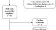
A systematic literature search of PubMed/MEDLINE and Embase databases was performed following PRISMA guidelines. The full-text articles methodological quality was independently assessed by four authors using the CASP-diagnostic study checklist.
Results
Out of 510 articles, 81 were selected of which 42 related to oncology. In early-recurrent prostate cancer (PSA range ≤ 0.5 and 0.5–2.0 ng/ml), PSMA-dPET has shown a significantly higher detection rate compared to aPET especially for smaller lesions. A higher image quality and lesion detectability was reported in [18F]FDG studies on lung cancer and on mixed oncological cohorts, where metabolic TNM upstaging occurred in up to 32% of cases compared to aPET. dPET technology was also found to improve the localization of in-transit metastases in melanoma, the staging of early oral squamous cell carcinoma, as well as the accuracy of [68 Ga]Ga-DOTA-TATE and 124I imaging in neuroendocrine tumors and thyroid cancer respectively. Although dPET sensitivity can provide better image quality in diagnostic and therapeutic (90Y-SIRT) applications, the possible higher rate of false positive findings (e.g., unspecific bone uptake at PSMA-1007), and SUVmax/radiomic-features variability should be considered. Main studies limitations included their retrospective nature, heterogeneity, and matched pair comparison design.
Conclusions
dPET has shown a diagnostic advantage over aPET in a variety of oncological settings, where the earlier and more accurate lesion localization and quantification could have relevant implications for optimal patient management.
期刊介绍:
Clinical and Translational Imaging is an international journal that publishes timely, up-to-date summaries on clinical practice and translational research and clinical applications of approved and experimental radiopharmaceuticals for diagnostic and therapeutic purposes. Coverage includes such topics as advanced preclinical evidence in the fields of physics, dosimetry, radiation biology and radiopharmacy with relevance to applications in human subjects. The journal benefits a readership of nuclear medicine practitioners and allied professionals involved in molecular imaging and therapy.
 求助内容:
求助内容: 应助结果提醒方式:


