{"title":"Exploring the Molecular Underpinnings of Skin Regeneration and Wound Healing: The Role of Renin Angiotensin.","authors":"Seyedeh Hoda Qoreishi, Mohammad Amin Khazeei Tabari, Mihnea-Alexandru Găman, Armaghan Kazeminejad","doi":"10.18502/ajmb.v16i3.15740","DOIUrl":null,"url":null,"abstract":"<p><p>The aim of this study is to review the role of renin-angiotensin in skin regeneration and wound healing with a focus on molecular mechanisms. Angiotensin receptor type 1 (AT1R) are abundant in the wounded area, and thus, lead to the activation of ERK, STAT1, and STAT3 which can lead to epidermal self-renewal. The expression of Renin Angiotensin System (RAS) components was significantly lower in wounds caused by burning, rather than intact skin, noting that RAS is involved in the re-epithelialization of skin. ERK, STAT and STAT3 are the targets of Ang II, indicating that RAS active components are involved in fibroblast, stem cells and keratinocyte migration. The effect of inhibiting the RAS on wound healing is context-dependent. On one hand, it is suggested that inhibiting RAS during this phase may slow down wound healing speed. On the other hand, studies have shown that RAS inhibition in this phase can lead to α-SMA activation, ultimately accelerating the wound healing process. Most of the investigations indicate that the inhibition of RAS with Angiotensin Receptor Blockers (ARBs) and Angiotensin Converting Enzyme (ACE) plays a significant role in tissue remodeling in the last phase of wound healing. It has been shown that the inhibition of RAS can inhibit scar formation and fibrosis through the downregulation of inflammatory and fibrogenic agents, such as TGF-β, SMAD2/3, and TAK1, PDGF-BB, and HSP47. To sum up, that local administration of RAS regulators might lead to less scar formation and inflammation in the last phase of wound closure.</p>","PeriodicalId":8669,"journal":{"name":"Avicenna journal of medical biotechnology","volume":"16 3","pages":"146-155"},"PeriodicalIF":0.0000,"publicationDate":"2024-07-01","publicationTypes":"Journal Article","fieldsOfStudy":null,"isOpenAccess":false,"openAccessPdf":"https://www.ncbi.nlm.nih.gov/pmc/articles/PMC11316511/pdf/","citationCount":"0","resultStr":null,"platform":"Semanticscholar","paperid":null,"PeriodicalName":"Avicenna journal of medical biotechnology","FirstCategoryId":"1085","ListUrlMain":"https://doi.org/10.18502/ajmb.v16i3.15740","RegionNum":0,"RegionCategory":null,"ArticlePicture":[],"TitleCN":null,"AbstractTextCN":null,"PMCID":null,"EPubDate":"","PubModel":"","JCR":"Q3","JCRName":"Biochemistry, Genetics and Molecular Biology","Score":null,"Total":0}
引用次数: 0
Abstract
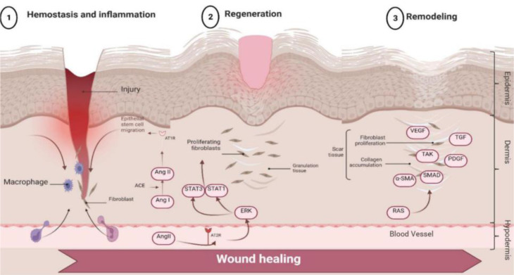
The aim of this study is to review the role of renin-angiotensin in skin regeneration and wound healing with a focus on molecular mechanisms. Angiotensin receptor type 1 (AT1R) are abundant in the wounded area, and thus, lead to the activation of ERK, STAT1, and STAT3 which can lead to epidermal self-renewal. The expression of Renin Angiotensin System (RAS) components was significantly lower in wounds caused by burning, rather than intact skin, noting that RAS is involved in the re-epithelialization of skin. ERK, STAT and STAT3 are the targets of Ang II, indicating that RAS active components are involved in fibroblast, stem cells and keratinocyte migration. The effect of inhibiting the RAS on wound healing is context-dependent. On one hand, it is suggested that inhibiting RAS during this phase may slow down wound healing speed. On the other hand, studies have shown that RAS inhibition in this phase can lead to α-SMA activation, ultimately accelerating the wound healing process. Most of the investigations indicate that the inhibition of RAS with Angiotensin Receptor Blockers (ARBs) and Angiotensin Converting Enzyme (ACE) plays a significant role in tissue remodeling in the last phase of wound healing. It has been shown that the inhibition of RAS can inhibit scar formation and fibrosis through the downregulation of inflammatory and fibrogenic agents, such as TGF-β, SMAD2/3, and TAK1, PDGF-BB, and HSP47. To sum up, that local administration of RAS regulators might lead to less scar formation and inflammation in the last phase of wound closure.

 求助内容:
求助内容: 应助结果提醒方式:


